|
||
|
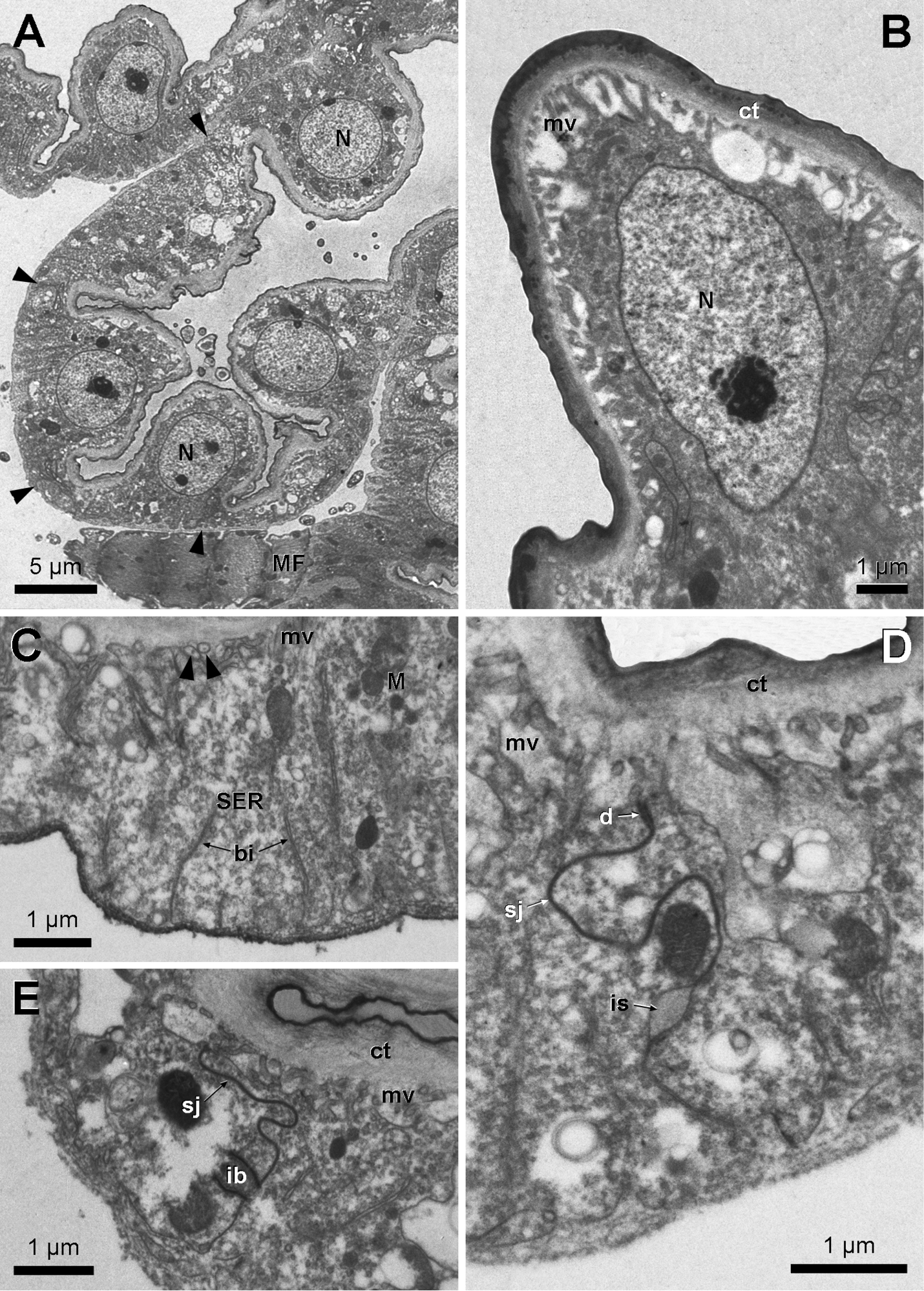
Electron micrographs of Dufour gland secretory cells in Myzinum sp.1. A Low magnification survey of crenellate epithelium, arrowheads indicate cell junctions B Cupola-like apical part of gland cell with nucleus (N) C Low lateral part of gland cell with apical microvilli (mv) and basal invaginations (bi). Arrowheads indicate extensions of smooth endoplasmic reticulum (SER) into microvilli D Detail of intercellular junction with apical desmosome (d), followed by septate junction (sj). Note intercellular space (is) wedged in between neighbouring cell walls. E. Occurrence of intercellular bridge (ib). ct: cuticle, M: mitochondria. |