|
||
|
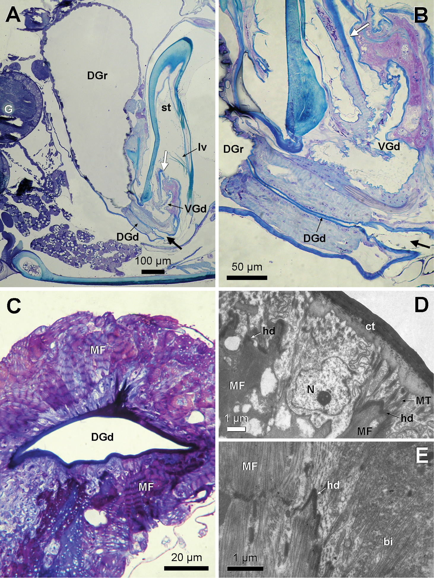
A Longitudinal semithin section through sting base region in Myzinum sp.2, showing Dufour gland opening ventrally of the sting base (black arrow), whereas the venom gland duct opens through the sting (white arrow) B Enlargement from A showing sting base region C Cross semithin section through Dufour gland duct in Myzinum sp.1, showing slit-like duct with attachment of dorsal and ventral muscle fibres (MF) D and E Electron micrographs of muscular attachments onto Dufour gland duct of Myzinum sp.1. Myofilaments of muscle fibres (MF) transmit their pulling force onto bundles of microtubules (MT) in the duct cells via hemidesmosomes (hd). bi: basal invaginations, DGd: Dufour gland duct, DGr: Dufour gland reservoir sac. G: ganglion, lv: lancet valves, N: nucleus, st: sting, VGd: venom gland duct. |