


(C) 2012 Jenő Papp. This is an open access article distributed under the terms of the Creative Commons Attribution License 3.0 (CC-BY), which permits unrestricted use, distribution, and reproduction in any medium, provided the original author and source are credited.
For reference, use of the paginated PDF or printed version of this article is recommended.
Descriptions are given of five new braconid species and one new genus from Colombia: Aspilota stigmalis sp. n., Synaldis cauca sp. n., Telmogarbus gen. n., Telmogarbus olivai sp. n. (all Alysiinae); Blacus (Tarpheion) latestigma sp. n. (Blacinae)and Pseudorhysipolis inaequalis sp. n. (Rhysipolinae). Types are deposited in the A. Humboldt Institute, Villa de Leyva, Boyacá, Bogota (Colombia). Critical remarks on the taxonomic position of the genus Synaldis are included. With 57 line drawings.
New species, new genus, type, description, taxonomy, neotropics
In the Neotropical braconid material, sent by Dr. M. J. Sharkey (University of Kentucky) to me for identification, seven specimens were found which proved to represent five new species, one species also a new genus. The five species were collected in Colombia with Malaise traps. The new species are as follows (in brackets the respective subfamily name): Aspilota stigmalis sp. n. (Alysiinae: Alysiini), Blacus (Tarpheion) latestigma sp. n. (Blacinae), Pseudorhysipolis inaequalis sp. n. (Rhysipolinae), Synaldis cauca sp. n. (Alysiinae: Alysiini) and Telmogarbus olivai gen. et sp. n. (Alysiinae: Alysiini). The type specimens are deposited in the Insect Collection, Alexander Humboldt Institute, Villa de Leyva, Boyacá, Bogota, Colombia; one paratype is in the Hungarian Natural History Museum, Budapest, Hungary.
Descriptions of the new taxaIn the descriptions abbreviations follow
Subfamily Alysiinae, tribe Alysiini
urn:lsid:zoobank.org:act:0F26E475-5F9E-4F96-8407-AC2DD4BED718
http://species-id.net/wiki/Aspilota_stigmalis
Figures 1–9COLOMBIA, Magdalena PNN Sierra Nevada de Santa Marta Bella Vista, 10°48'N, 73°39'W, 1500 m, Malaise trap, 1–15 June 2001, leg. J. Cantillo. – Holotype is in good condition: (1) glued on a card point by the right mesopleuron, (2) left antenna missing ultimate or 19th antennomere (17th flagellomere), (3) right pair of wings less visible owing to mounting and to the apically creased fore wing.
The species name stigmalis refers to the large pairs of spiracles on the propodeum and first tergite (Figs 5, 9).
Body 3 mm long. Antenna as long as head, mesosoma and tergites 1–2 combined, with 19 antennomeres. Scape twice as long as broad apically and somewhat belly, first flagellomere 1.2 times as long as second, second flagellomere somewhat thicker than first, first flagellomere 3.7 times and second 2.5 times as long as broad (Fig. 1). Head in dorsal view transverse, almost 1.9 times as broad (between temples) as long (between compound eye and temple), temple slightly swollen, eye almost 1.3 times as long as temple (Fig. 2). Eye in lateral view 1.7 times as high as wide and just wider than gena, gena evenly broad beyond eye. Mandible along its lower margin 1.8 times as long as broad between upper and lower teeth, both teeth rounded (Fig. 3). Tentorial pit fairly large and extending to lower part of eye (Fig. 4). Head polished.
Mesosoma in lateral view stout, somewhat longer than high, polished. Notauli short, restricted to anterior declivous part of mesoscutum and finely crenulate. Pronope missing. Precoxal suture short, crenulate, medially on mesopleuron. Propodeum areolate, spiracles large, otherwise propodeum smooth and shiny, on its upper corner foveolate depressed (Fig. 5). Hind femur 3.8 times as long as broad distally (Fig. 6). Hind tibia slightly longer than hind tarsus.
Fore wing as long as body. Pterostigma linear (Fig. 7), r long, 3.5 times as long as width of pterostigma. Second submarginal cell long, 3–SR 2.3 times as long as 2–SR, r–m as long as r. First discal cell: 1–M 1.9 times as long as 1–SR–M (Fig. 8).
First tergite (Fig. 9) long, twice as long as posteriorly broad, spiracles large and at middle of tergite, pair of basal keels reaching spiracles, hind half of tergite striolate. Tergites 2–3 fully fused (i.e. border between tergites indistinct) and together with further tergites polished. Ovipositor sheath as long as mid tibia.
Scape and pedicel yellow, flagellum darkening brown. Head and mesosoma chestnut dark brown. Mouthparts whitish. First tergite brownish yellow, tergites 2–3 brownish, remaining tergites chestnut dark brown. Legs pale yellow. Tibiae apically and tarsi faintly light brownish. Wings hyaline, pterostigma brown, veins light brown.
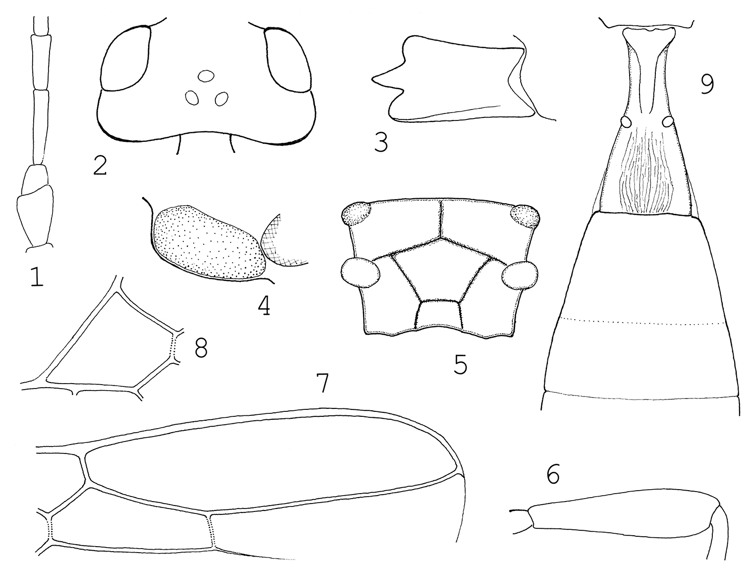
Aspilota stigmalis sp. n.: 1 scape, pedicel and flagellomeres 1–2 2 head in dorsal view 3 mandible 4 paraclypeal pit 5 propodeum 6 hind femur 7 distal part of right fore wing 8 first discal cell of fore wing 9 tergites 1–3.
unknown.
Colombia.
The new species, Aspilota stigmalis, is nearest to Aspilota phyllotomae Fischer (
| 1(2) | Propodeum granulo-rugulose and not areolate, spiracles of propodeum and first tergite small, i.e. usual in size. Antenna with 14–15 antennomeres, flagellomeres 1–2 equal in length (each about three times as long as broad) and equally thick. Eye in dorsal view just shorter (cf. Abb. 33 in |
Aspilota phyllotomae Fischer, 1970 |
| 2(1) | Propodeum areolate, otherwise smooth and shiny, spiracles of propodeum and first tergite large (Figs 5, 9). Antenna with 19 antennomeres, first flagellomere 1.2 times as long as second flagellomere, second flagellomere somewhat thicker than first flagellomere (Fig. 1). Eye in dorsal view almost 1.3 times as long as temple (Fig. 2). Fore wing: r more than two times as long as width of pterostigma, 3–SR 2.3 times as long as 2–SR, SR1 2.25 times as long as 3–SR (Fig. 7). Prosternum and tegula brown. ♀: 3 mm. – Colombia | Aspilota stigmalis sp. n. |
urn:lsid:zoobank.org:act:78AEA130-5EF3-4FD2-A9BF-0415E0DCAFBC
http://species-id.net/wiki/Synaldis_cauca
Figures 10–16COLOMBIA, Valle del Cauca, PNN Farallones de Cali, Anchicaya, 3°26'S, 76°48'W, 900 m, Malaise trap, 1 August – 10 October 2000, leg. S. Sarria. – Holotype is in good condition: (1) glued on a card point by its right mesopleuron, (2) right antenna broken, with 15 antennomeres, (3) right fore wing creased apico-posteriorly.
The new species is named after the type locality, ”Cauca”.
Body 2.6 mm long. Antenna as long as body and with 21 antennomeres. First flagellomere three times as long as broad apically, further flagellomeres gradually shortening and indistinctly attenuating so that penultimate flagellomere 2.5 times as long as broad. Head in dorsal view less transverse or subcubic (Fig. 10), 1.7 times as broad as long, eye almost 2.9 times as long as temple, temple rounded. Ocelli medium-sized, OOL almost three times as long as POL. Eye in lateral view nearly 1.5 times as high as wide and nearly 1.6 times as wide as temple, temple beyond eye evenly widened (Fig. 11, see arrows). Lower tooth of mandible somewhat small, mandible twice as long as broad between upper and lower teeth (Fig. 12). Paraclypeal pit short, i.e. distance between pit and eye as long as length of paraclypeal pit itself (Fig. 13). Maxillary palp one-sixth longer than height of head.
Mesosoma in lateral view stout, somewhat longer than high, polished. Mesoscutal dimple before prescutellar furrow. Precoxal suture medially on mesopleuron, crenulate. Propodeum with medio-longitudinal carina and with areolation (areola basalis, etc., Fig. 14). Hind femur 4.1 times as long as broad distally (Fig. 15). Hind tibia and tarsus equal in length. Hind basitarsus as long as tarsomeres 2–3 combined.
Fore wing: venation ”Synaldis-form”, i.e. 2–SR missing hence first and second submarginal cells confluent; r + 3–SR as long as SR1, CU1a issuing from middle of outer side of subdiscal cell.
First tergite (Fig. 16) 2.8 times as long as broad posteriorly, moderately broadening posteriorly. Pair of keels merging into fine striation; spiracles close beyond middle of tergite. Tergites 2–3 fused and as long as first tergite, together with further tergites polished. Ovipositor sheath as long as mid tibia.
Scape and pedicel brownish yellow, flagellum darkening brown. Head, mesosoma and first tergite brownish black, rest of metasoma brown. Mandible and mouthparts yellow. Tegula brownish yellow. Legs yellow, hind tarsus greyish fumous. Wings hyaline, pterostigma and veins opaque brownish yellowish.
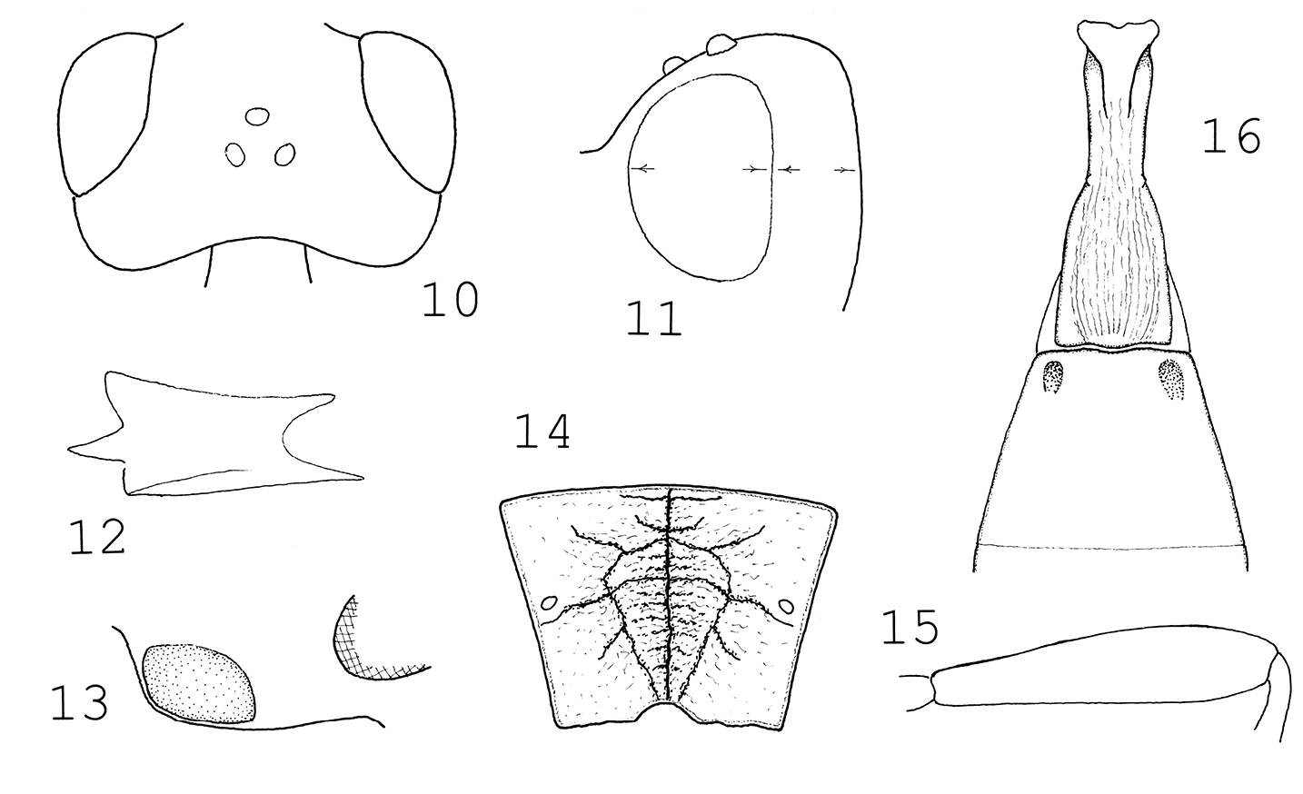
Synaldis cauca sp. n.: 10 head in dorsal view 11 head in lateral view 12 mandible 13 paraclypeal pit 14 propodeum 15 hind femur 16 tergites 1–2.
unknown.
Colombia.
The new species, Synaldis cauca, is near to Synaldis acutidens Fischer as both have a mandible with three spiky teeth, SR1 more than twice as long as r + 3–SR combined and dark bodies; the two species are distinguished as follows (Synaldis acutidens is known by its original description:
| 1(2) | Head in dorsal view 1.5 times as broad as long; eye as long as temple. Propodeum polished and with medio-longitudinal carina, spiracles fairly large. Antenna with 16–18 antennomeres. Hind femur 3.5 times as long as broad distally (Abb. 2 in |
Synaldis acutidens Fischer, 1967 |
| 2(1) | Head in dorsal view 1.7 times as boad as long; eye almost 2.9 times as long as temple (Fig. 10). Propodeum with medio-longitudinal carina and with areolation, spiracles small (Fig. 14). Antenna with 21 antennomeres. Hind femur 4.1 times as long as broad distally (Fig. 15). First tergite 2.6 times as long as broad, widening less posteriorly (Fig. 16). Lower tooth of mandible somewhat small (Fig. 12). Head, mesosoma and first tergite brownish black, metasoma brown. ♀: 2.6 mm. – Colombia | Synaldis cauca sp. n. |
The single distinctive generic feature of the genus Synaldis Foerster that differentiates it from Aspilota Foerster (the confluent first and second submarginal cells of the fore wing, or absence of vein 2–SR) has been questioned since more than a century.
The above short essay is but a viewpoint in the taxonomic treatment of the genus Synaldis. My conception is expounded in a traditional form – the morphological data matrix and molecular analysis will, presumably, unambiguously solve this taxonomic problem.
urn:lsid:zoobank.org:act:6F9EFAFB-3F27-4A38-BFE9-C84E3B673E0A
http://species-id.net/wiki/Telmogarbus
Figures 20–24, 33–40Telmogarbus olivai gen. et sp. n. (monobasic and present designation).
The new genus receives the fantasy name, Telmogarbus.
Body gracile. Antenna filiform, flagellomeres very long. Head in dorsal view transverse, eye distinctly longer than temple (Fig. 36). Mandible with four teeth, fourth tooth between first and second teeth (Fig. 20, see arrow). Maxillary palp very long, about twice as long as height of head. Face and clypeus much wider than high. Paraclypeal pit short, i.e. not reaching compound eye (Fig. 35). Ocelli medium-sized, elliptic. Pronope small, distinct. Notauli evenly deep, meeting behind, finely crenulate; mesoscutal dimple missing. Prescutellar furrow wide, with a few crenulae. Axille with pair of lamelliform excrescences (Fig. 23). Metanotum medially with thin, long spine curving posteriorly (Fig. 24).Propodeum polished, carinate (Fig. 38). Precoxal suture distinct, crenulate. Epicnemial carina present. Legs long and thin. Fore wing as long as body. Pterostigma widening medially, second submarginal cell long: 3–SR clearly longer than 2–SR, m–cu interstitial (Fig. 21). First tergite petiolate, beyond first tergite metasoma narrow, tergites polished, tergites 2–3 fused (Fig. 33). Hypopygium small, ovipositor sheath short. Ground colour of body yellow to (light) brown. Wings hyaline.
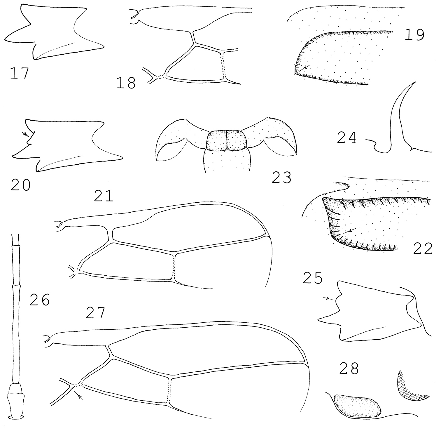
17–19 Cratospila circe (Haliday): 17 mandible 18 pterostigma and first submarginal cell of right fore wing 19 mesoscutum in lateral view (its declivous lateral part). 20–24 Telmogarbus olivai gen. et sp. n.: 20 mandible 21 distal part of right fore wing 22 mesoscutum in lateral view (its declivous lateral part) 23 axilla with pair of excrescenses 24 metanotal spine in lateral view. 25–28 Kritscherysia longimembrum Fischer: 25 mandible 26 scape, pedicel and flagellomeres 1–2 27 distal part of right fore wing 28 paraclypeal pit.
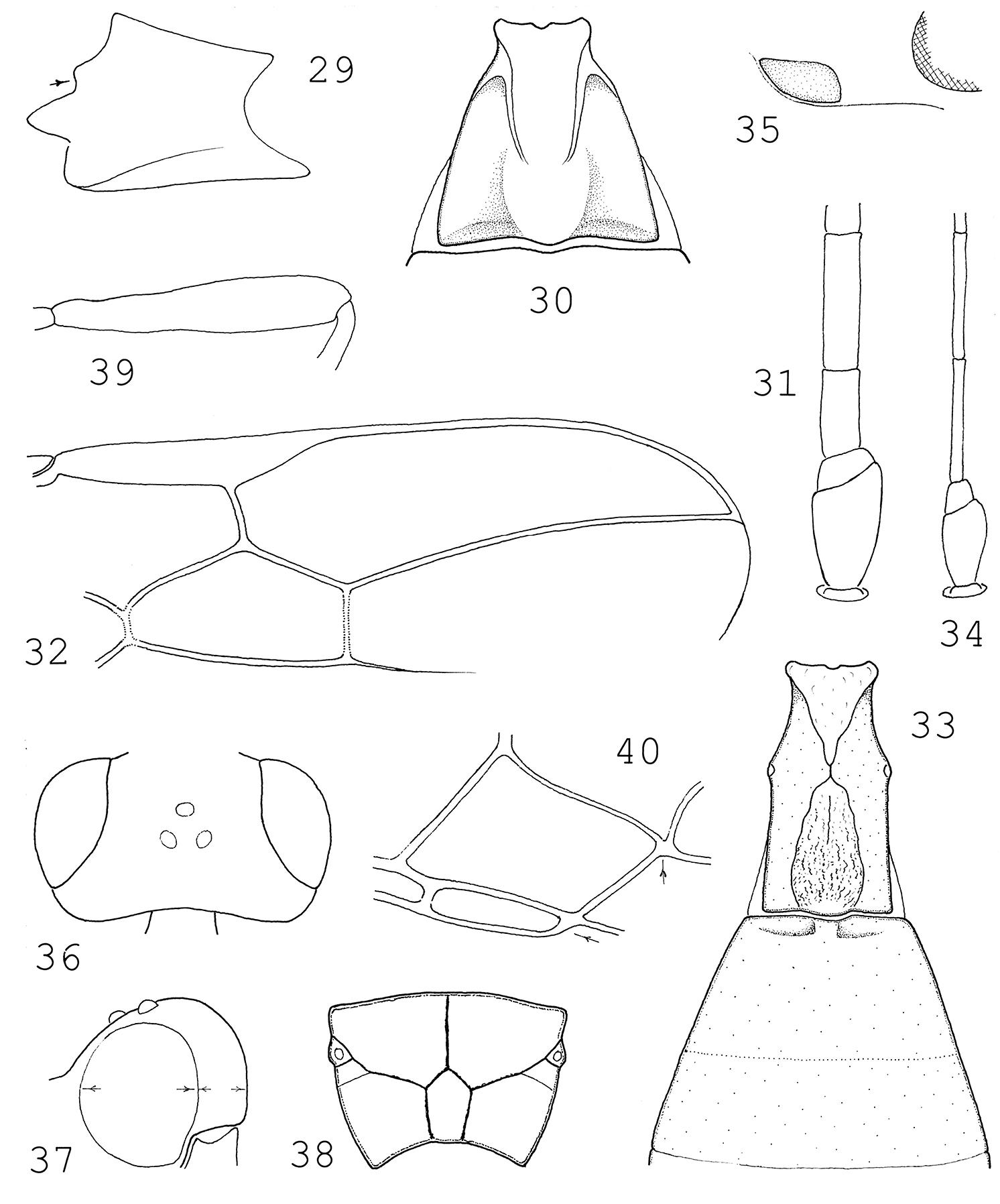
29–32 Gnathopleura cariosa Marsh: 29 mandible 30 first tergite 31 scape, pedicel and flagellomeres 1–2 32 distal part of right fore wing. 33–40 Telmogarbus olivai gen. et sp. n.: 33 tergites 1–3 34 scape, pedicel and flagellomeres 1–2 35 paraclypeal pit 36 head in dorsal view 37 head in lateral view 38 propodeum 39 hind femur 40 first discal and first subdiscal cells.
The new genus is near to the Palaearctic genus Kritscherysia Fischer (
1.) Distinction between Kritscherysia and Telmogarbus: Common features of the two genera are the pair of more or less lamelliform excrescenses laterally from the prescutellar furrow (or on axillae) (Fig. 23; Abb. 54 in
| 1(2) | Metanotum without spine. Mandible with convexity between teeth 1 and 2 (”Interkalarwölbung” |
Kritscherysia Fischer, 1993 |
| 2(1) | Metanotum with spine (Fig. 24). Mandible with a fourth tooth between teeth 1 and 2 (Fig. 20, see arrow). Flagellomeres 1 and 2 equal in length (Fig. 34). Fore wing: r issuing from middle of pterostigma (Fig. 21), n. rec. interstitial (Fig. 40, see vertical arrow). Paraclypeal pit short, its length as long as distance between pit and compound eye (Fig. 35). One species in the Neotropical Region | Telmogarbus gen. n. |
2.) Distinction between Gnathopleura and Telmogarbus: The new genus runs to Gnathopleura Fischer in Wharton’s (1997: 88–98) identification key to the alysiine genera of the Neotropical Region, their common feature is the fourth tooth between teeth 1 and 2 (Figs 20, 29, see arrows); the distinction between them is presented here.
| 1(2) | Mandible strong and broad, 1.2–1.4 times as long as broad between upper and lower teeth (Fig. 29). First tergite as long as to 1.2 times as long as broad posteriorly, strongly widening posteriorly (Fig. 30). Flagellomeres thick, first flagellomere 2 to 3 times as long as broad, second flagellomere variably longer / as long as / shorter than first flagellomere (Fig. 31). Fore wing: 3–SR at most as long as 2–SR or shorter, i.e. second submarginal cell short (Fig. 32). Wings dark, infumate. – Twelve described species in the Neotropical Region ( |
Gnathopleura Fischer, 1975 |
| 2(1) | Mandible usual in size, 1.8 times as long as broad between upper and lower teeth (Fig. 20). First tergite 1.8 times as long as broad posteriorly, subparallel-sided (Fig. 33). Flagellum thin, first flagellomere 10 times as long as broad, second flagellomere as long as first flagellomere (Fig. 34). Fore wing: 3–SR 1.5 times as long as 2–SR, i.e. second submarginal cell long (Fig. 21). Wings hyaline. – One species in the Neotropical Region | Telmogarbus gen. n. |
3.) Distinction between Cratospila and Telmogarbus: disregarding the number of mandibular teeth, the new genus will run to Cratospila Foerster (
| 1(2) | Mandible with three teeth (Fig. 17). Fore wing: 3–SR shorter than (or at most as long as) 2–SR, i.e. second submarginal cell short; pterostigma with r issuing distal to its middle (Fig. 18; Fig. 83 in |
Cratospila Foerster, 1862 |
| 2(1) | Mandible with four teeth, fourth tooth between first and second teeth (Fig. 20, see arrow). Fore wing: 3–SR 1.5 times as long as 2–SR, i.e. second submarginal cell long; pterostigma with r issuing from its middle (Fig. 21). Notauli turning into crenulate lateral margin of mesoscutum (Fig. 22, see arrow). Axilla with lamelliform excrescense (Fig. 23); metanotum with spine (Fig. 24). One species in the Neotropical region | Telmogarbus gen. n. |
Taxonomic remark. The three genera related to the new genus Telmogarbus are known to me by the following material: 1.) Cratospila by females and males of Cratospila circe Haliday; 2.) Gnathopleura by a few Neotopical species; 3.) Kritscherysia by two male paratypes of Kritscherysia longimembrum Fischerand the original description.
urn:lsid:zoobank.org:act:198BCB2B-3030-4C87-B2D3-D944CFA2E478
http://species-id.net/wiki/Telmogarbus_olivai
Figures 20–24, 33–40COLOMBIA, Nariño R. N., La Planada Parcela Olga, 1°15'N, 78°15'W, Malaise trap, holotype: 16 June – 2 July 2000 and paratype: 16 March – 2 April 2001, leg. G. Oliva. Holotype is in fairly good condition: (1) glued on a card point by lower part of mesopleuron, ;(2) right flagellum broken, antenna with 19 antennomeres; (3) missing: right middle leg, right fore leg (except coxa) and tarsomeres 2–4 of left hind leg; (4) left fore wing apically missing beyond pterostigma. Paratype is also in fairly good condition: (1) glued on card point by meso- and metapleura; (2) left flagellum apically broken, antenna with 22 antennomeres; (3) missing tarsi of right fore and hind legs.
The new species is dedicated to its collector, Mr. G. Oliva.
Female: Body 3.6 mm long. Antenna long, twice as long as body, with 33 antennomeres. Flagellomeres very long, flagellomeres 1–2 equal in length and ten times as long as broad apically (Fig. 34), subsequent flagellomeres slightly shortening so that penultimate flagellomere four times as long as broad. Head in dorsal view transverse (Fig. 36), 1.8 times as broad as long, eye 4.5 times as long as temple, temple rounded, occiput weakly excavate. OOL somewhat more than twice as long as POL. Eye in lateral view 1.25 times as high as wide and 2.4 times as wide as temple, temple beyond eye evenly wide (Fig. 37). Mandible 1.8 times as long below as broad between upper and lower teeth, fourth tooth between upper and middle teeth, every tooth pointed (Fig. 20). Paraclypeal pit as long as distance between pit and compound eye (Fig. 35). Segments of maxillary palp long, palp about 1.5 times as long as height of head. Head polished.
Mesosoma in lateral view stout, 1.2 times as long as high, polished. Pronope present. Crenulate notauli turning down to crenulated margin of mesoscutum, i.e. notauli not extending onto declivous anterior part of mesoscutum (Fig. 22, see arrow). Notauli meeting posteriorly (before prescutellar furrow) and here dimple missing. Prescutellar furrow subcrenulate, laterally from furrow (or on axillae) with pair of lamelliform excrescenses (Fig. 23). Metanotum medially with fairly long and somewhat posteriorly curved spine (Fig. 24). Propodeum areolate, areolae polished, spiracles before middle of propodeum, carination strong (Fig. 38). Precoxal suture distinct, restricted to middle of mesopleuron, crenulate, hind margin of mesopleuron almost smooth to subcrenulate; epicnemial carina present. Legs long. Hind femur 5.5 times as long as broad distally (Fig. 39). Hind tibia almost 1.3 times as long as hind tarsus.
Pterostigma (Fig. 21) 6.6 times as long as wide and r issuing from its middle, r short, shorter than width of pterostigma. Second submarginal cell long, 3–SR 1.6 times as long as 2–SR, n. rec. interstitial, SR1 1.6 times as long as 3–SR and reaching tip of wing. First subdiscal cell narrowing distally and closed (Fig. 40, see horizontal arrow), CU1a interstitial (Fig. 40, see vertical arrow).
First tergite (Fig. 33) 1.8 times as long as broad behind, weakly broadening posteriorly, spiracles before middle of tergite; pair of basal keels meeting at spiracle and posteriorly diverging, reaching hind end of tergite. Hind half of tergite medially between keels longitudinally substriate, otherwise polished. Tergites 2–3 fused and as long as first tergite, together with further tergites polished (Fig. 33). Ovipositor sheath as long as first tergite or hind basitarsus + half of second tarsomere.
Ground colour of body yellow to brownish yellow. Scape and pedicel brownish yellow. Flagellum brown to dark brown, flagellomeres 19–25 white. Head brownish yellow, vertex brown. Mandible, mouthparts, palps pale yellow. Prothorax browish yellow, mesoscutum, scutellum and mesosternum brown. Mesopleuron and propodeum blackish brown. Tergites brown, sternites yellow. Legs yellow, coxae and trochanters lemon yellow. Wings hyaline, pterostigma and veins yellowish.
Male paratype. Similar to female holotype. Body 3.5 mm long. Antenna with 31 antennomeres. Head in dorsal view 1.8 times as broad as long. Ptero-stigma wide, 5.7 times as long as wide. Dark colour somewhat more extensive.
Unknown.
Colombia.
Subfamily Blacinae
urn:lsid:zoobank.org:act:AF970EB5-9078-4098-AE34-B6B8D1B0AD93
http://species-id.net/wiki/Blacus_latestigma
Figures 41–50COLOMBIA, Amazonas, PNN Amacayacu San Martin, 150 m, 3°23'S, 70°6'W, Malaise trap, leg. B. Amado. Holotype is in good condition: (1) glued on a card point by the right metapleuron, (2) right femur + tibia in glue, hardely visible, (3) left hind leg on the right side owing to the mounting.
The name latestigma refers to the wide pterostigma.
Body 2.5 mm long. Antenna somewhat longer than body and with 25 antennomeres. First flagellomere six times as long as broad apically and 1.5 times as long as second flagellomere, subsequent flagellomeres shortening so that penultimate flagellomere twice as long as broad. Head in dorsal view (Fig. 41) distinctly twice as broad as long, eye almost four times as long as temple, temple receding. Occiput carinate. Ocelli elliptic, OOL twice as long as POL. Malar suture present. Maxillary palp somewhat longer than height of head. Eye in lateral view 1.3 times as high as wide. Head polished.
Mesosoma in lateral view 1.4 times as long as high. Notauli distinct, smooth. Precoxal suture short with weak crenulae. Lateral carina of scutellum missing. Propodeum rugose (like Fig. 101 in van
Fore wing almost as long as body. Pterostigma wide (Fig. 44), 2.6 times as long as wide, r issuing somewhat distal to middle, r 0.8 times as long as width of pterostigma. 2–SR nearly 1.6 times as long as r, 3–SR + SR1 slightly bent and reaching tip of wing, 2–M short. First subdiscal cell open distally (Fig. 45, see arrow). Hind wing: cu–a slightly proximal to middle of vein M+CU + 1–M (Fig. 46, see arrow).
First tergite long and hardly broadening posteriorly (Fig. 47), 2.2 times as long as broad posteriorly, pair of keels distinct, finely striate. Second tergite almost 1.9 times as broad posteriorly as long, anteriorly strio-rugulose. Further tergites polished. Hypopygium fairly large, ovipositor sheath downcurved, somewhat longer than hind tibia.
Scape, pedicel and flagellomeres 1–2 straw yellow, rest of flagellum brown. Head brownish yellow, occiput brown, mouthparts whitish. Mesosoma blackish brown, tegula brown. Legs yellow, coxae and trochanters straw yellow. First tergite blackish brown, further tergites brown, anterior sternites whitish. Wings hyaline, pterostigma brown, parastigma whitish, veins opaque brown.
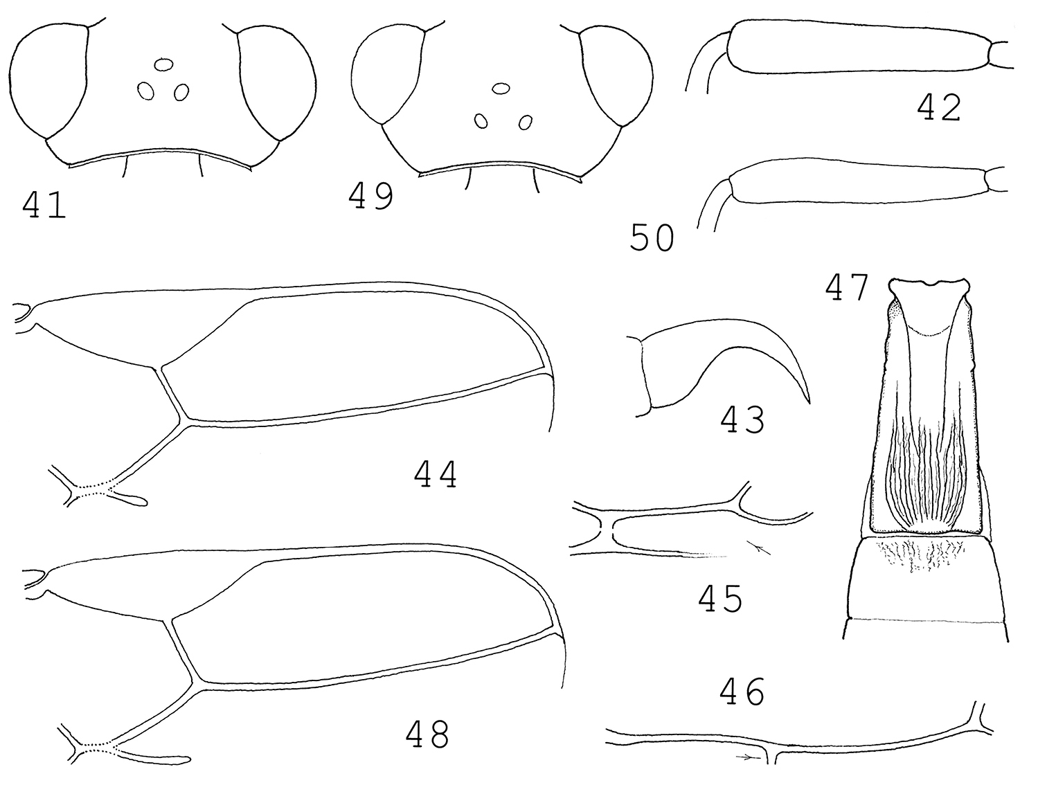
41–47 Blacus (Tarpheion) latestigma sp. n.:41 head in dorsal view 42 hind femur 43 claw 44 distal part of right fore wing 45 first subdiscal cell 46 hind wing: M+CU + 1–M+cu–a 47 tergites 1–2. 48–50 Blacus (Tarpheion) erugatus van Achterberg: 48 distal part of right fore wing 49 head in dorsal view 50 hind femur.
unknown.
Colombia.
The new species, Blacus (Tarpheion) latestigma, runs to Blacus (Tarpheion) erugatus van Achterberg with the help of van Achterberg’s keys (1976: 186, 1988: 140–141); the two species differ from each other by a few features.
| 1(2) | Fore wing: pterostigma narrower, 3–3.3(3.6) times as long as wide, r as long as width of pterostigma, 2–M long (Fig. 48). Eye in dorsal view 2.3 times as long as temple, temple slightly less receding, head in dorsal view slightly less transverse: 1.9 times as broad as long (Fig. 49). Hind femur 5.5–6.2 times as long broad distally (Fig. 50). First tergite more broadening posteriorly (Fig. 101 in van |
Blacus (Tarpheion) erugatus van Achterberg, 1976 |
| 2(1) | Fore wing: pterostigma wide, 2.6 times as long as wide, r 0.8 times as long as width of pterostigma, 2–M short (Fig. 44). Eye in dorsal view 3.8 times as long as temple, temple slightly more receding, head in dorsal view clearly transverse, twice as broad as long (Fig. 41). Hind femur five times as long as broad distally (Fig. 42). First tergite hardly broadening posteriorly (Fig. 46). Antenna with 25 antennomeres. Ground colour of mesosoma blackish brown, head brownish yellow, occiput brown. ♀: 2.5 mm. – Colombia | Blacus (Tarpheion) latestigma sp. n. |
Taxonomic remark. Blacus (Tarpheion) erugatus is known to the author by a pair of female specimens, their data: (a) Costa Rica, Puntar, Golfo Dulce, 24 km W Piedras-Blancas, 200 m, June–August 1989, leg. Hanson; (b) Costa Rica, Carthago Pr., La Cangreja, 1950 m, October 1991, leg. Hanson. Both females identified by S.R. Shaw in 1999 and in the Hungarian Natural History Museum (Budapest) by exchange of material. The species is new to the fauna of Costa Rica.
Subfamily Rhysipolinae
urn:lsid:zoobank.org:act:8076B0BB-A892-4EF2-B203-345925A26426
http://species-id.net/wiki/Pseudorhysipolis_inaequalis
Figures 51–57COLOMBIA, Amazonas, PNN Amacayacu Mata-Mata, Malaise trap, 150 m, 2–15 October 2001, leg. D. Chota. M 2239. Holotype is in good condition: (1) glued on a card point by its right meso- and metapleura, (2) left antenna broken, flagellum with 20 flagellomeres. Paratype is in fairly good condition: (1) glued on a card point by its right mesopleuron, (2) both antennae broken, right antenna with 16 and left antenna with 13 antennomeres, (3) right fore wing somewhat creased distally. Holotype in Boyacá, Colombia; one female paratype in Museum Budapest, Hym. Typ. No. 12021.
The species name inaequalis refers to the unequal lengths of 3–SR and 2–M of the fore wing (Fig. 55).
Body 3.3 mm long. Antenna (right one) somewhat longer than body and with 30 antennomeres. First flagellomere a little longer than second, first flagellomere 3.5 times as long as broad apically, subsequent flagellomeres shortening and attenuating so that penultimate flagellomere 3.6 times as long as broad. Head in dorsal view transverse (Fig. 51), almost 1.7 times as broad as long, eye fairly large: 7.5 times as long as temple, temple receding. Ocelli small, elliptic, OOL twice as long as POL. Eye in lateral view 1.3 times as high as wide and 5.4 times as wide as temple, beyond eye evenly wide (Fig. 52). Horizontal diameter of oral opening twice as long as shortest distance between opening and eye. Maxillary palp as long as height of head. Occipital carina completely removed from hypostomal carina. Face and gena smooth and shiny, vertex subgranulose, subshiny to matt.
Mesosoma in lateral view nearly twice as long as high. Mesoscutum, scutellum and propodeum granulose, otherwise mesosoma smooth and shiny. Pronope absent. Notauli complete, deep, smooth. Prescutellar furrow crenulate. Precoxal suture weakly distinct, smooth. Propodeum granulose, with a medio-longitudinal carina dividing posteriorly, polished anterior to and around spiracles (Fig. 53). Hind femur 3.8 times as long as broad medially (Fig. 54). Inner apex of hind tibia with comb-like dense bristles (cf. Fig. 15 in
Fore wing as long as body. Pterostigma (Fig. 55) four times as long as wide and r issuing from its middle, somewhat longer than width of pterostigma (12:10). Second submarginal cell long, 3–SR twice as long as 2–SR; SR1 faintly bent, slightly more than twice as long as 3–SR and reaching tip of wing. Vein r, 2–SR and m–cu equal in length. First subdiscal cell long, distally closed (Fig. 56, see arrow).
First tergite (Fig. 57) 1.5 times as long as broad posteriorly, weakly broadening posteriorly, dorsope distinct, pair of basal keels ending before middle of tergite, domed median part of tergite granulose. Second tergite quadrate, a little broader behind than long medially; tergites 2–4 largely weakly sclerotized or membranous (Fig. 57). Ovipositor sheath long, as long as mid tibia.
Scape and pedicel ochre, flagellum brown. Ground colour of head and mesosoma ochre; scutellum, propodeum and tergites brown. Mouthparts yellow, palps straw yellow. First tergite dark brown. Legs yellow, coxae and trochanters 1–2 straw yellow. Wings hyaline, pterostigma brownish, basally and apically yellow, veins yellowish to brownish.
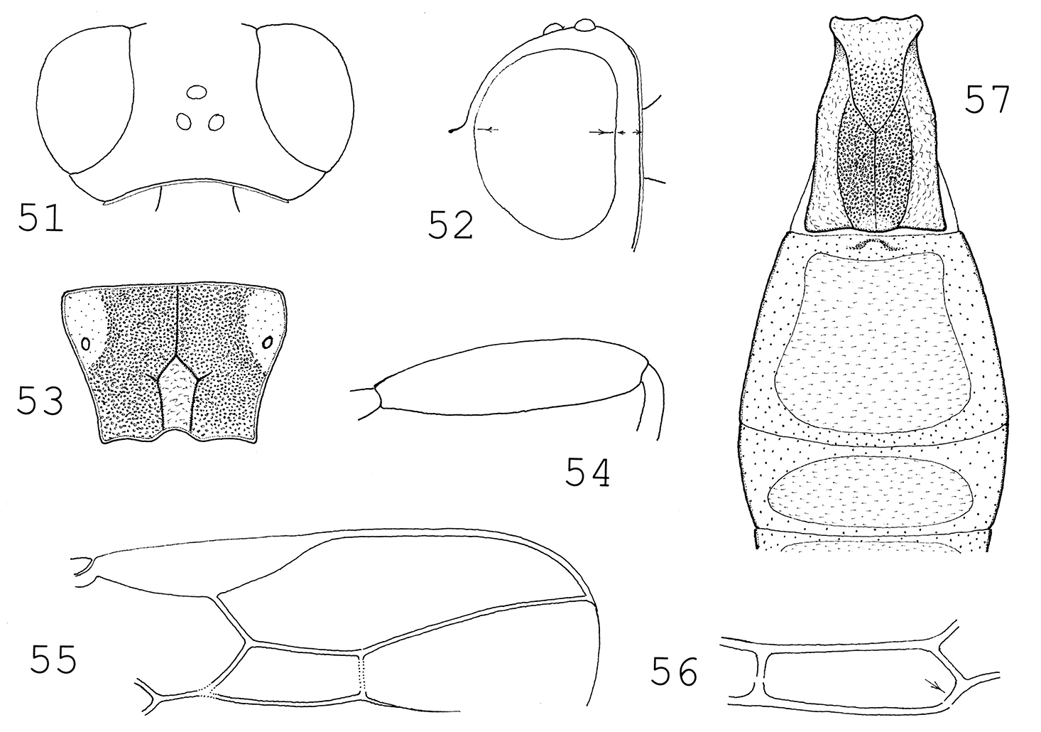
Pseudorhysipolis inaequalis sp. n.: 51 head in dorsal view 52 head in lateral view 53 propodeum 54 hind femur 55 distal part of right fore wing 56 first discal cell 57 tergites 1–3.
Similar to the female holotype. Body 3.3 mm long. Head somewhat dark ochre.
unknown.
Colombia.
The new species, Pseudorhysipolis inaequalis, runs to Pseudorhysipolis notaulicus van Achterberg & Penteado-Dias in
| 1(2) | Fore wing: 3–SR and 2–M equal in length, i.e. second submarginal cell rectangular (Fig. 12 in |
Pseudorhysipolis notaulicus van Achterberg & Penteado-Dias, 2002 |
| 2(1) | Fore wing: 3–SR 0.6 times as long as 2–M, i.e. second submarginal cell usual in form (Fig. 55). Eye in dorsal view 7.5 times as long as temple (Fig. 51). First tergite 1.5 times as long as broad posteriorly (Fig. 57). Hind femur 3.8 times as long as broad (Fig. 54). Pterostigma basally yellow, mesoscutum ochre. ♀: 3.3 mm. – Colombia | Pseudorhysipolis inaequalis sp. n. |
I am much indebted to the curators for their kindness to promote my taxonomic work by loaning type and other braconid material: Dr M. Fischer (Wien), Dr M. J. Sharkey (Lexington) and Dr S. R. Shaw (Laramie). Also my sincere thank should go to Dr. G. Broad (London) as peer referee considerably improved the content as well as the linguistic style of my paper.