|
||
|
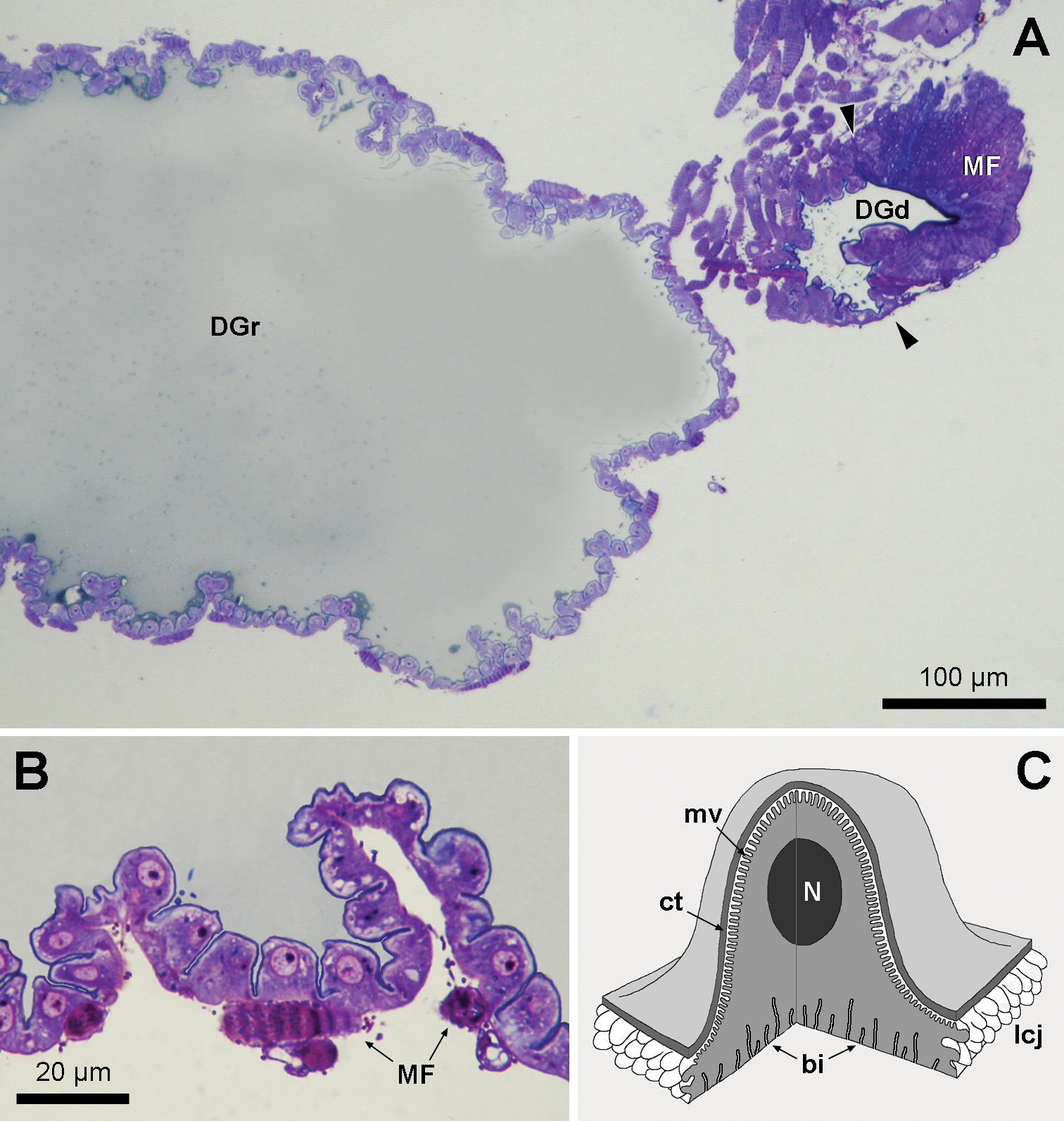
A Semithin section through the Dufour gland reservoir sac (DGr) and duct region (DGd) in Myzinum sp.1. The arrowheads indicate the transition between the reservoir and duct region B Detail of crenellate epithelial lining of reservoir sac with strands of surrounding muscle fibres (MF) C Schematical view of cupola-like shape of gland cell. bi: basal invaginations, ct: cuticle, lcj: lateral cell junction, mv: microvilli, N: nucleus. |