|
||
|
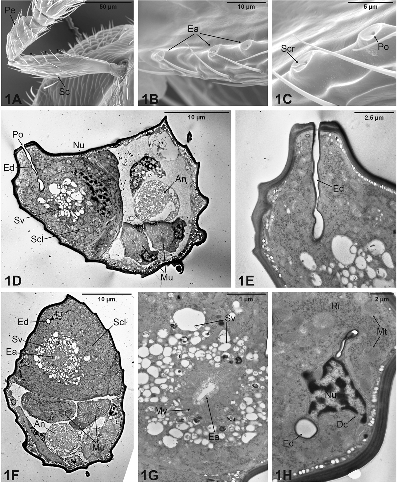
Structure of the male scape in Aphelinus varipes. A–C Scanning electron microscope images showing in A the modified male scape (Sc) with the presence of a ventral carina on which three specialized structures (arrowheads) are observed B detail of the carina revealing three elevated areas (Ea) C close up of the previous in which a single apical pore (Po) per Ea is clearly visible, as well as secretion (Scr) oozing from the pore itself. D–H Transmission electron microscope images showing internal ultrastructural features of the male scape D cross section of the scape taken through one of the elevated areas, showing the secretory cell (Scl) that occupies about half of the scape internal volume; Scl has a large nucleus (Nu) located basally, a central cluster of electron-lucid secretory vesicles (Sv) and a straight evacuating duct (Ed) connected with the external pore (Po); the rest of the scape volume is occupied by muscles (Mu) and the antennal nerve (An) E close-up view of the previous image, showing the cuticular evacuating duct (Ed) running straight towards the external pore F cross section of the scape taken in a different view, showing the large secretory cell (Scl) with a centrally positioned end apparatus (Ea) surrounded by numerous secretory vesicles (Sv); the evacuating duct (Ed), muscles (Mu), and antennal nerve (An) can be seen G detail of the end apparatus (Ea), which appears perforated and surrounded by microvilli (Mv); secretory vesicles (Sv) are above H detail of the duct cell (Dc) characterised by a very reduced cytoplasm and a small nucleus (Nu); the duct cell is surrounded by the cytoplasm of the secretory cell, that reveals the presence of ribosomes (Ri) and mitochondria (Mt); the evacuating duct (Ed) is visible in cross section. Scale bars: 50 µm (A); 10 µm (B, D, F); 5 µm (C); 2.5 µm (E); 1 µm (G); 2 µm (H). |