|
||
|
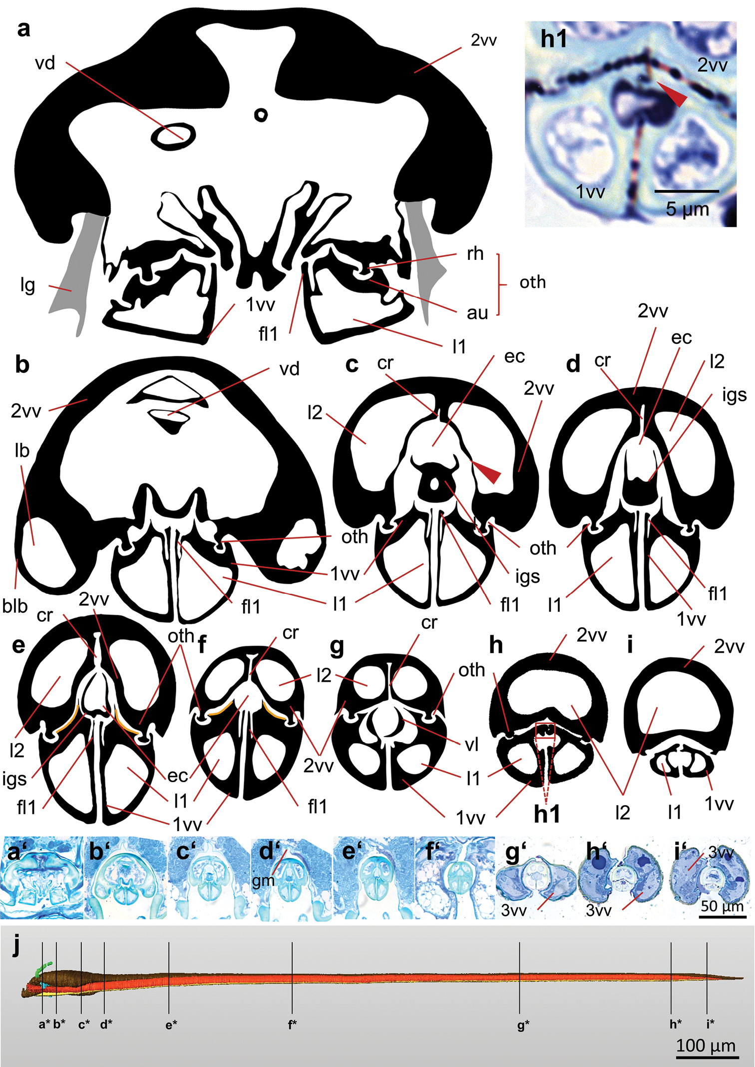
(next page) Cross sections through the terebra of Habrobracon hebetor (from proximal to distal); schematic drawings of the 1st and 2nd valvula (a–i) based on the light microscopic images of the presented semithin sections (a’–i’ 600 nm; stained with Stevenel’s blue). The drawings are of the same size ratio. The 2nd valvulae possesses, in the proximal region, two lumina that merge into one in the most distal region (h–i). The bulbs (b) and the valvilli (g) are visible. The orange lines (in e, f) mark the position of the distally pointing ctenoid structures on the dorsal surfaces of the 1st valvulae, which are in close contact with the ventral surface of the 2nd valvula. The genital membrane connects the dorsal margins of the 2nd valvifers (b’–h’) c fine cuticular structures arise from the dorsal and ventral parts of the 2nd valvula and define the lumina of the bulbs (arrow) h1 olistheter-like interlocking system connecting the medial surfaces of the apices of the paired 1st valvulae (arrow). Final segmented 3D reconstruction based on a semithin section series (600 nm thickness) a*–i* position of each single section marked on the final 3D reconstruction of the terebra. Abbreviations: 1vv = 1st valvula; 2vv = 2nd valvula; 3vv = 3rd valvula; au = aulax; blb = bulb; cr = longitudinal crack of 2nd valvula; ec = egg canal; fl1 = longitudinal flap of the 1st valvula; gm = genital membrane; igs = internal guiding structure; l1 = lumen of 1st valvula; l2 = lumen of 2nd valvula; lb = lumen of the bulb; lg = ligament; oth = olistheter; rh = rhachis; vd = duct of the venom gland; vl = valvillus. |