


(C) 2012 Michael J. Sharkey. This is an open access article distributed under the terms of the Creative Commons Attribution License 3.0 (CC-BY), which permits unrestricted use, distribution, and reproduction in any medium, provided the original author and source are credited.
For reference, use of the paginated PDF or printed version of this article is recommended.
Based on a phylogenetic analysis, the limits of Therophilus (Hymenoptera: Braconidae: Agathidinae) are redefined and restricted to a small proportion of the previously included species. Those species belonging to the world fauna are listed and the species from Thailand are revised. Forty-four species are assigned to the genus including 11 new species, i.e. Therophilus anuchati, Therophilus apichati, Therophilus areeluckae, Therophilus boonthami, Therophilus chiangmaiensis, Therophilus kwanuiae, Therophilus songrami, Therophilus sukpengae, Therophilus wannai, Therophilus wongchaii, Therophilus wongwani. A dichotomous key to species is presented; links to an electronic interactive key and to distribution maps are also included.
Thailand, Insecta, identification key, taxonomy, systematics
Agathidinae is a moderately large subfamily of Braconidae with 1, 069 described species worldwide and 246 in the Oriental Region (
As part of the inventory of Thai insects, we ran 3 Malaise traps at each of 30 different localities throughout Thailand from 2007-2010, comprising approximately 90 Malaise traps. The specimens dealt with here are primarily from these traps.
Species concepts are based on morphological data and 28S rDNA data. Regions D2-D3 of 28S rDNA (roughly 560 base pairs) were sequenced using the following primers: 28SD2hymF 5’ - AGAGAGAGTTCAAGAGTACGTG - 3’ and 28SD3hymR 5’ - TAGTTCACCATCTTTCGGGTC - 3’. Sequences were edited using Geneious Pro v4.7.5 (
Phenetic and phylogenetic trees were constructed using neighbor-joining (NJ), maximum parsimony (MP) and Bayesian methods. MP was performed using TNT (
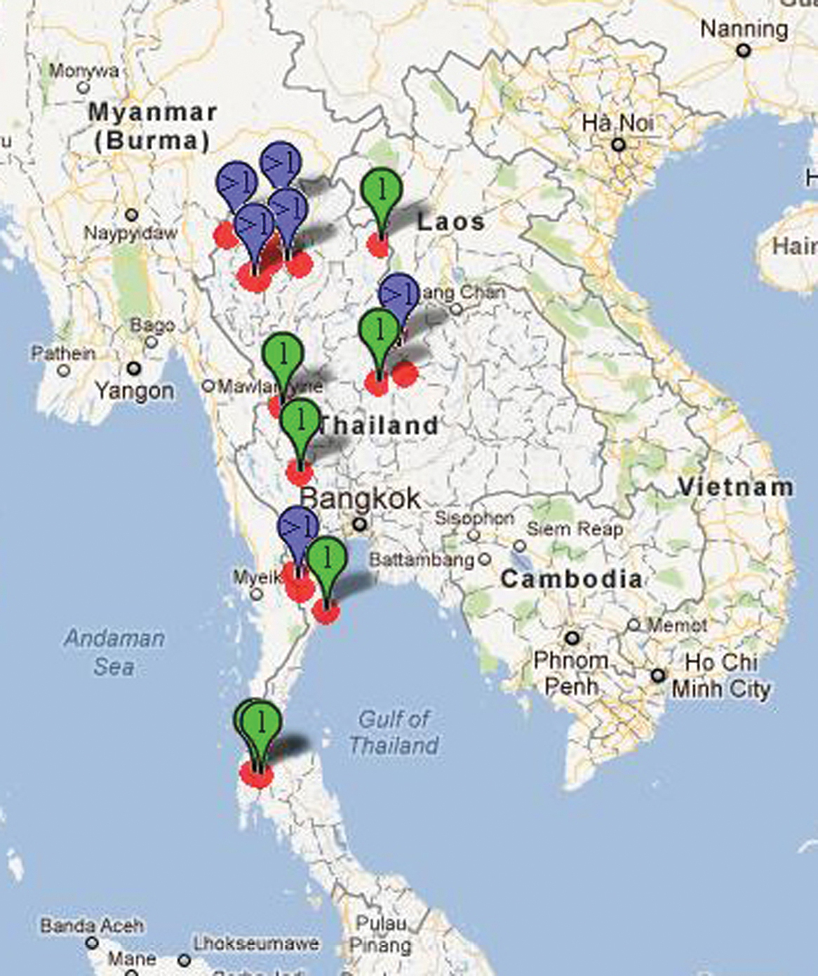
Map showing Therophilus collection sites in Thailand.
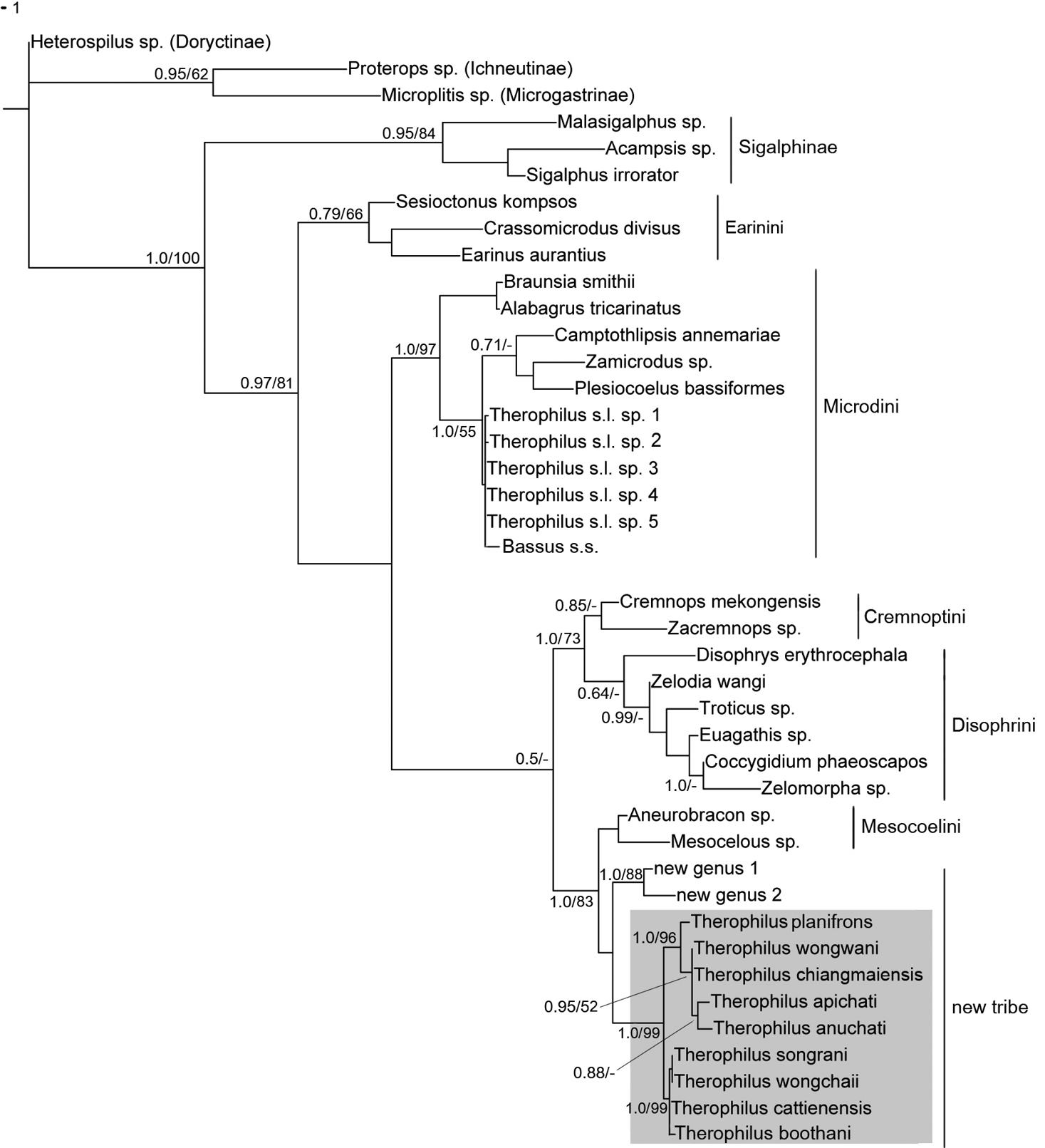
NJ phylogram based on 28S rDNA. Where Bayesian and parsimony analyses agreed with the NJ tree, branch support values are included in the figure, i.e., Bayesian posterior probabilities/parsimony bootstrap (values below 0.5 posterior probability and/or 50% bootstrap support were not recorded on the tree). Therophilus s.s. highlighted in grey box.
The dichotomous key, descriptions, and the interactive key (Appendices 1–3) were generated using DELTA Editor (
Morphological terms follow
Morphological terms used in this revision were matched to the Hymenoptera Anatomy Ontology (HAO,
All 14 species found in Thailand are treated with a diagnosis and distributional data. They are illustrated with color photos using a JVC digital camera mounted on a Leica MZ16 microscope and Automontage® stacking software. Distributional data are listed for all species and a Google map via Berkeley Mapper is included for all species. The descriptions are of the holotype and variation is given in parentheses.
The source files for the keys, descriptions, illustrations, DNA sequence and distributional data are all freely available to future researchers who may wish to build on these data. Distribution data, pdf’s of non-copywrite references, images, notes, and host and type information can be found by searching Taxabank (a combined specimen and taxonomic database; http://purl.org/taxabank). Codes beginning with an “H” and followed by numbers are unique identifiers used for specimens in the Sharkey lab at the University of Kentucky, and in the specimen database TaxaBank (e.g., H235). All sequences have been deposited in the GenBank database.
Abbreviations used for specimen depositories are as follows:HIC Hymenoptera Institute Collection, University of Kentucky, Department of Entomology, Lexington, Kentucky, USA.
QSBG Queen Sirikit Botanic Gardens, Chiang Mai, Thailand.
RMNH NCB Naturalis Collection [formerly Rijksmuseum van Natuurlijke Historie], Leiden, Netherlands.
IRSNB Institut Royal des Sciences Naturelles de Belgique, Brussels, Belgium.
Results PhylogenyRecently the polyphyletic generic concept, Bassus, was divided into four genera, i.e., Bassus s.s., a small monophyletic group confined to the Old World, Lytopylus, a large monophyletic group with a world-wide distribution, Neothlipsis, a small New World genus and Therophilus, a polyphyletic dumping ground for the remaining species. Here we refine the concept of Therophilus so that it is monophyletic. However, this leaves those species that do not fit the concept without a correct generic placement. We refer to these as Therophilus s.l. in this treatment, and they will be treated in a separate publication (Sharkey et al. in prep).
The phylogenetic tree in figure 2 shows that Therophilus in the strict sense, and two new genera, are far removed from the majority of the species of Therophilus sensu latu. The clade containing Therophilus s.s. was referred to as an unnamed new tribe in
Within Therophilus s.s., there are two distinct clades that may deserve generic status when more is known of their biology and diversity. All members of the lower clade, in figure 2, which includes Therophilus cattienensis, are stout and mostly yellow-orange, e.g., all have predominantly yellow-orange heads. Therophilus conspicuous Wesmael, the type species, belongs in this group. Members of the clade containing Therophilus chiangmaiensis are more gracile and melanic.
Microdus (Therophilus) conspicuous [Lectotype ♀ IRSNB, examined]
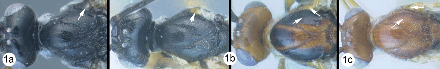
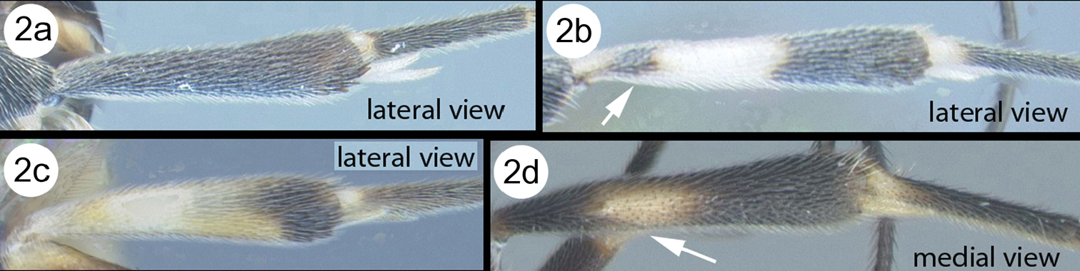
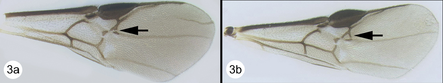
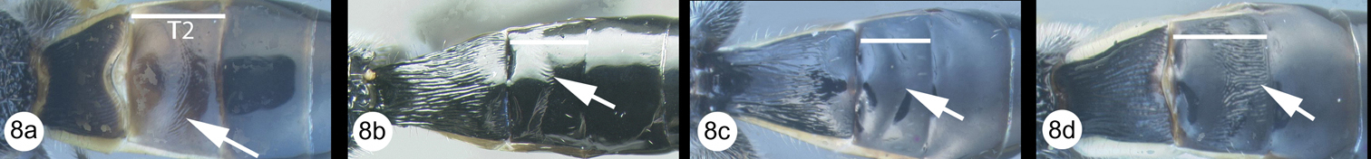
There is neither one character nor a specific combination of characters that distinguishes members of Therophilus from all other agathidines. It is easily separated from members of Cremnoptini and Disophrini by the shape of the tarsal claws, i.e., simple with a basal lobe in Therophilus, versus cleft claws in members of the two aforementioned tribes. Most members of Therophilus have the following combination of diagnostic characters: Apical abscissa of R of fore wing weak at midlength and bent towards apex of wing; interantennal space with two (usually weak and short) longitudinal ridges or with a median keel; postscutellar depression present; cubitus of hind wing strong and tubular, at least basally; subbasal cell of hind wing angled apically at the point from which the cubitus emanates; sclerite between hind coxal cavities and metasomal foramen incomplete or narrow. A combination of characters that distinguishes almost all species of Oriental Therophilus is: Interantennal space with longitudinal groove (Figs. 3c, 6c), sometimes weak (Fig. 8c); posteroscutellar depression present (Fig. 8f); notauli sculptured with pits (Fig. 3f).
Head. Lateral carina on frons absent (Fig. 3c); interantennal space with longitudinal groove (Fig. 3c, 6c); gena not extended ventroposteriorly into sharp prominence (Fig. 5e); gena lacking sharp angle posteriad eye; labial palpus with three or four segments, third segment much reduced or absent; apical antennomere acute but lacking nipple-like process.
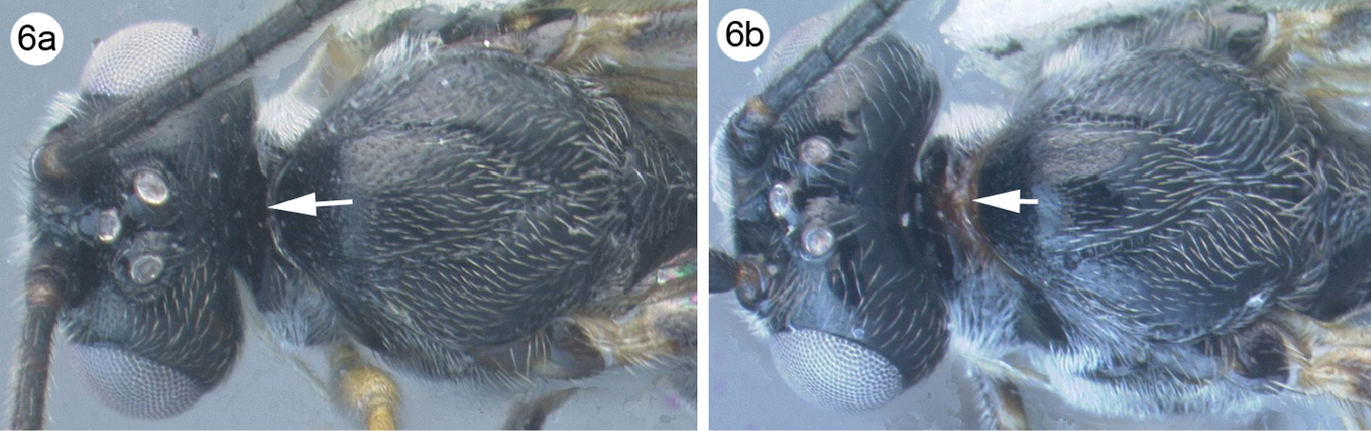
Mesosoma. Propleuron lacking a projection at mid height; notauli impressed and pitted, at least in part (Fig. 3f); posteroscutellar depression present (Fig. 8f) (rarely absent); propodeum from rugose to areolate-rugose (Fig. 7g); sclerite between hind coxal cavities and metasomal foramen narrow, sometimes incomplete.
Legs.Fore tibia lacking pegs, fore tarsal claws with basal lobe; mid tibia with apical and medial pegs; hind tibia with apical pegs.
Wings (Figs. 3b, 4b). Fore wing RS + M vein incomplete; second submarginal cell triangular; fore wing 3RSb decurved, weak at midlength; hind wing r and r-m crossveins absent; hind wing CUb present and strong, tubular at least basally.
Metasoma. MT1 with longitudinal striations, lacking dominant pair of longitudinal carinae (Fig. 4f); MT2 from smooth to striate, usually with some longitudinal striae and weak transverse striae in first transverse depression; MT3 smooth (Fig. 4f); ovipositor as long as or longer than metasoma (Fig. 4a).
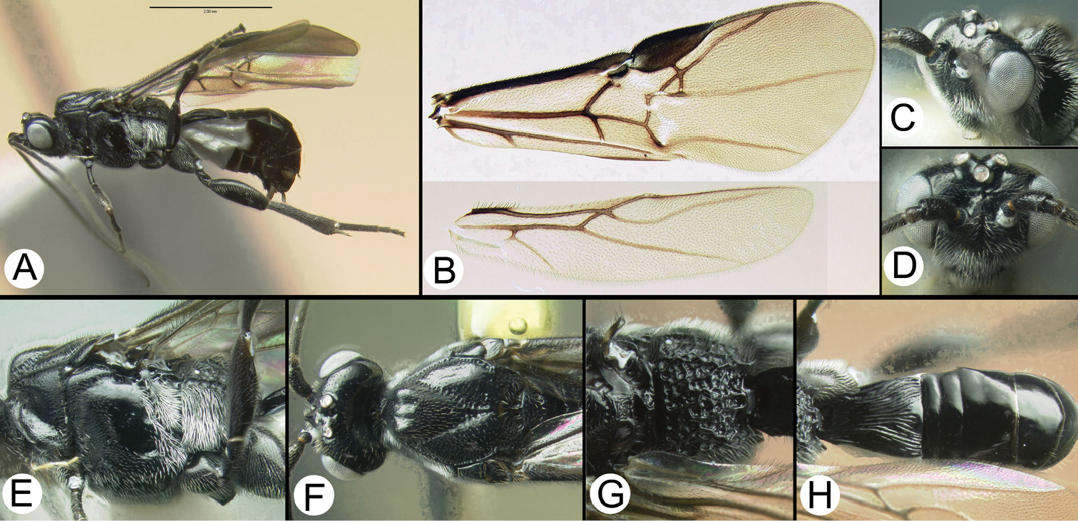
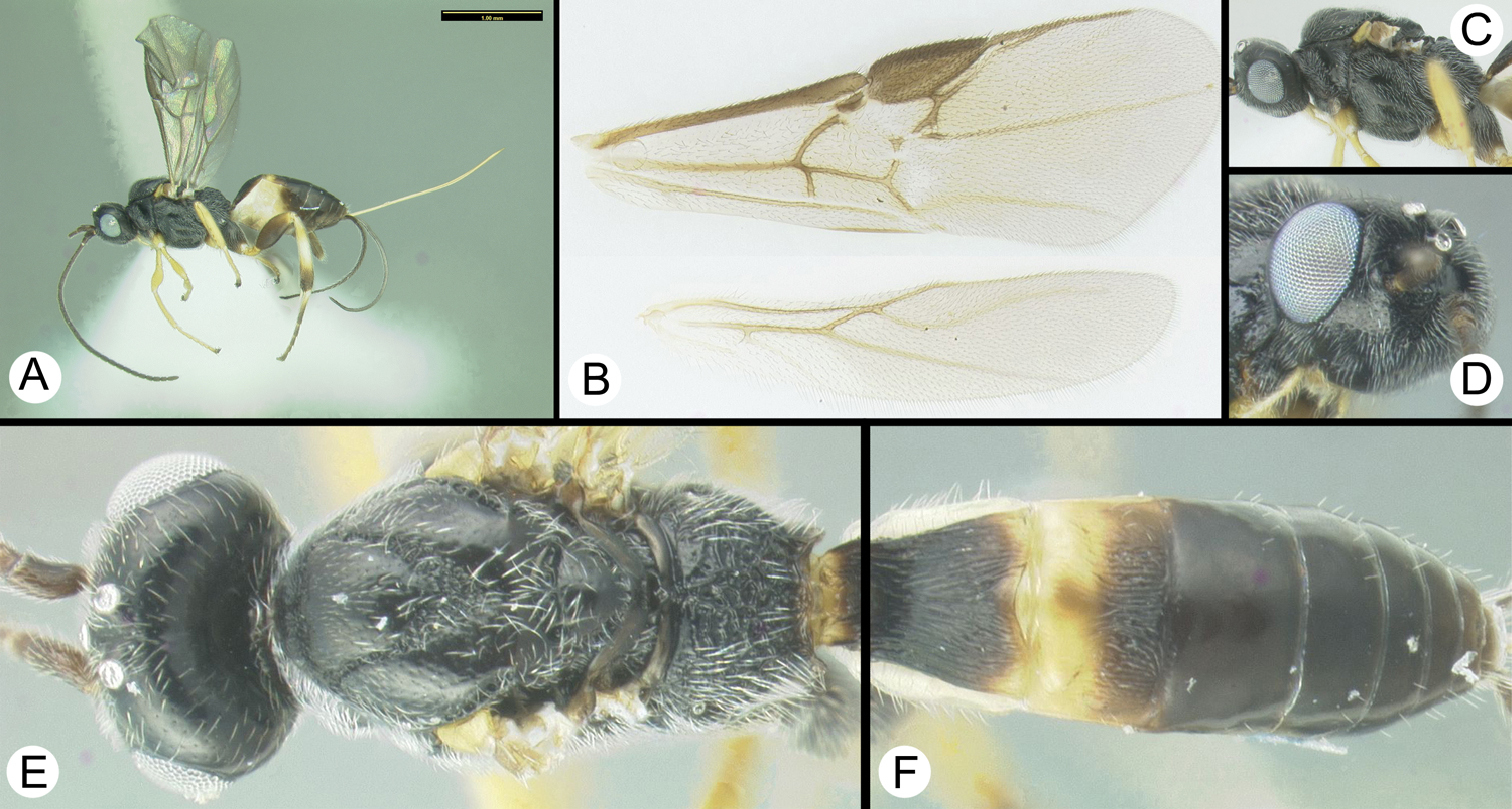
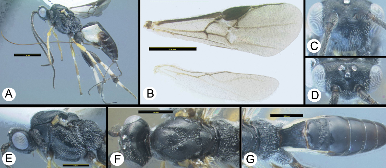
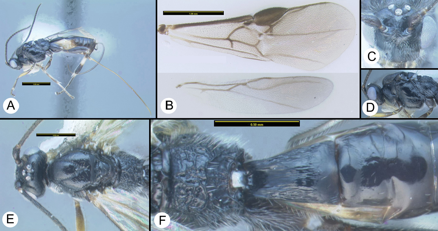
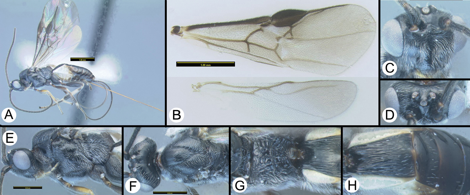
Therophilus anuchati sp. n. a lateral habitus b Wings c dorsolateral head d anterodorsal head e lateral mesosoma f dorsal head and mesosoma g dorsal propodeum h dorsal metasoma.
Including the twelve described here, there are 44 described species known to the senior author. The following 13 species were included in Therophilus at the time of this publication: Therophilus antipoda Ashmead, 1900, Therophilus arcuatus Reinhard, 1867, Therophilus cattienensis van Achterberg & Long, 2010, Therophilus cingulipes Nees, 1812, Therophilus clausthalianus Ratzeburg, 1844, Therophilus conspicuus Wesmael, 1837, Therophilus crenulisulcatus van Achterberg & Long, 2010, Therophilus levisoma van Achterberg & Long, 2010, Therophilus planifrons van Achterberg & Long, 2010, Therophilus rugosiferus van Achterberg & Long, 2010, Therophilus similis (Bhat & Gupta, 1977), Therophilus stephensae Stevens, 2011, Therophilus tumidulus (Nees, 1812).
The remainder are here transferred to Therophilus: Bassus arthurellus
According to
Worldwide, with more diversity in subtropical and tropical areas.
| 1 | a. Tegula black, concolorous with mesoscutum | 2 |
| b. Tegula yellow, contrasting with predominantly black mesoscutum | 8 | |
| c. Tegula yellow or orange, similar in color to predominantly orange or yellow mesoscutum | 12 | |
 |
||
| 2(1) | a. Hind tibia entirely melanic (check medial surface) | Therophilus anuchati Sharkey sp. n. |
| b. Hind tibia largely pale, melanic apically and with a subbasal melanic band or lateral spot | 3 | |
| c. Hind tibia mostly pale, melanic apically only | Therophilus kwanuiae Sharkey sp. n. | |
| d. Hind tibia mostly melanic with pale coloration at midlength at least medially | Therophilus chiangmaiensis Sharkey sp. n. | |
 |
||
| 3(2) | a. 2nd submarginal cell reduced to a small dot, petiole longer than cell is high | 4 |
| b. 2nd submarginal cell larger, cell height subequal to petiole length | 6 | |
 |
||
| 4(3) | a. Fore tarsus mostly or entirely melanic | 5 |
| b. Fore tarsus mostly or entirely pale | Therophilus kwanuiae Sharkey sp. n. | |
 |
||
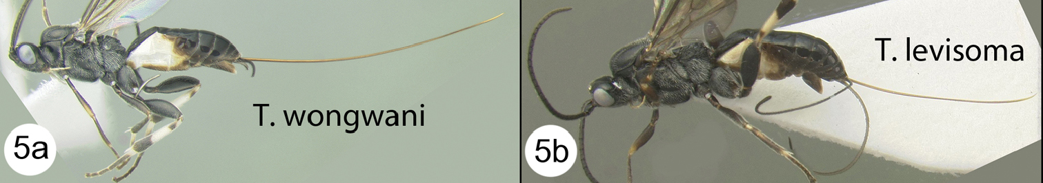
| 5(4) | a. Exposed portion of ovipositor distinctly longer than body | Therophilus wongwani Sharkey sp. n. |
| b. Exposed portion of ovipositor slightly shorter than body | Therophilus levisoma van Achterberg & Long | |
 |
||
| 6(3) | a. Pronotum entirely melanic | 7 |
| b. Pronotum mostly melanic but pale dorsomedially | Therophilus wannai Sharkey sp. n. | |
 |
||
| 7(6) | a. Posterior half of MT2 smooth | Therophilus crenulisulcatus van Achterberg & Long |
| b. Posterior half of MT2 longitudinally striate, at least medially | Therophilus sukpengae Sharkey sp. n. | |
 |
||
| 8(1) | a. MT2 mostly smooth with transverse and/or diagonal striae in and/or near transverse depression | Therophilus wongchaii Sharkey sp. n. |
| b. MT2 mostly smooth with short longitudinal striae restricted to transverse depression… | Therophilus planifrons van Achterberg & Long | |
| c. MT2 entirely smooth | 9 | |
| d. MT2 smooth in most of anterior half anteriad transverse groove, longitudinally striate in transverse groove and area posteriad transverse groove, at least medially. | 10 | |
 |
||
| 9(8) | a. MT2 entirely melanic | Therophilus planifrons van Achterberg & Long |
| b. MT2 entirely or almost entirely pale | Therophilus boonthami Sharkey sp. n. | |
 |
||
| 10(8) | a. MT2 entirely melanic | 11 |
| b. MT2 pale in anterior half, melanic posteriorly | Therophilus apichati Sharkey sp. n. | |
 |
||
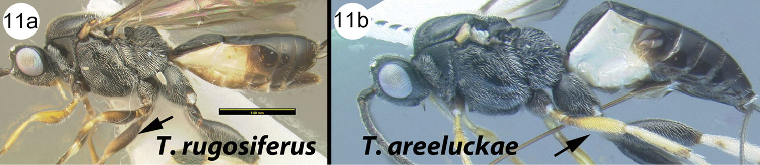
| 11(10) | a. Mid femur mostly or entirely melanic, usually pale distally | Therophilus rugosiferus van Achterberg & Long |
| b. Mid femur entirely or mostly pale, usually melanic at extreme base | Therophilus areeluckae Sharkey sp. n. | |
 |
||
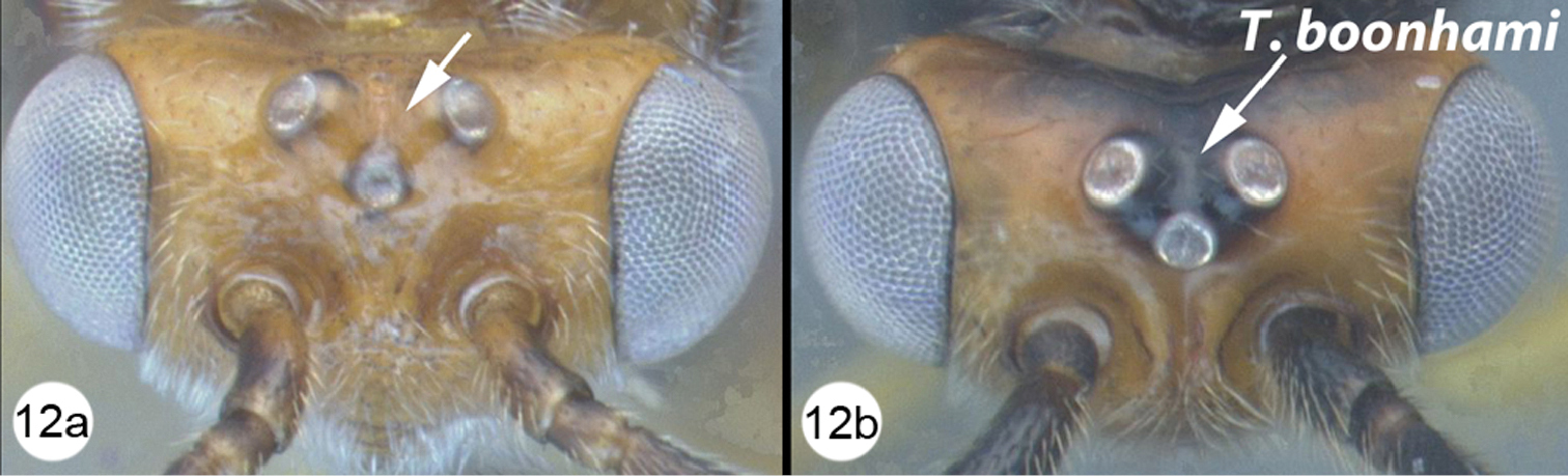
| 12(1) | a. Ocellar triangle pale, concolorous with remainder of vertex | 13 |
| b. Ocellar triangle melanic, contrasting with most of vertex | Therophilus boonthami Sharkey sp. n. | |
 |
||
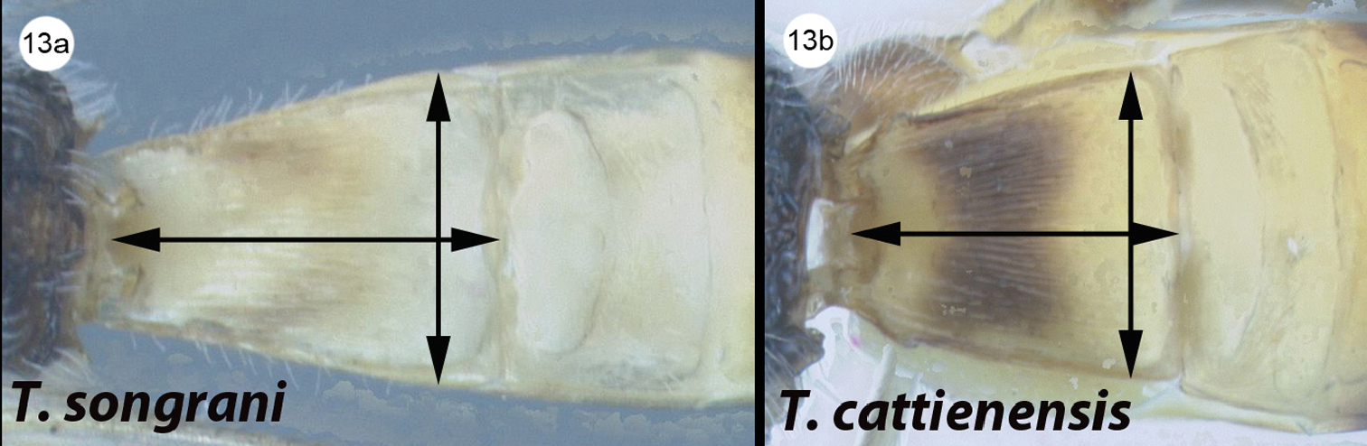
| 13(12) | a. MT1 length distinctly longer than apical width | Therophilus songrani Sharkey sp. n. |
| b. MT1 length only slightly longer than apical width | Therophilus cattienensis van Achterberg & Long | |
 |
||
urn:lsid:zoobank.org:act:134DEB0E-168C-4B81-94C4-A82CC11346DA
http://species-id.net/wiki/Therophilus_anuchati
Figure 3Ocellar triangle melanic, concolorous with remainder of vertex. Hind tibia entirely melanic. Strong transverse carinae between the hind coxal cavities and a wide sclerite between the hind coxal and metasomal foramina. Strong, sharply declivous longitudinal flange between antenna; hind wing CUb strong and long; median lobe of mesoscutum sharply sloping anteriorly.
Body length. 5.2 mm.
Head. Space between antennal insertions with a well-developed keel that is sharply declivous posteriorly, dorsal surface of keel with a shallow longitudinal groove. Number of flagellomeres 32. Posterior surface of scutellum completely rugose, posterior scutellar depression not distinct.
Mesosoma. Number of pegs on mid tibia = 4. Number of pegs on hind tibia = 9. Sclerite between metasoma and hind coxa wide with a high ridge along most or all of its length. Basal lobe of hind tarsal claw longer than high, not sharply declivous. Length-width of hind femur 1.0/0.342 = 2.9. 2nd submarginal cell large, cell height subequal to petiole length. Hind wing vein Cub emanates from near mid length of apical margin of subbasal cell, Cub long and strong. Point of notauli intersection heavily sculptured over a wide area with a median longitudinal ridge. Median lobe of mesoscutum bulging and sharply declivous anteriorly. Metapleuron with dense mat of white setae.
Metasoma. MT1 length distinctly longer than apical width. MT1 with widely-spaced longitudinal striae, lacking microsculpture between striae, and with and two pairs of distinctly stronger striae (carinae). MT1 distinctly wider apically than basally. Ratio of widest point of MT1 to narrowest point 0.61/0.411 = 1.5. Length-width ratio of MT1 0.8/0.61 = 1.3. MT2 entirely smooth.
Color. Mostly black or dark melanic except for dense white pilosity on metapleuron and white on anterior lateral tergites and sternites, more apical leg segments lighter, tending towards light brown or dark yellow, all apical spurs white, mandible and palpi mostly yellow, fore wing lightly infuscate, stigma brown with a small pale patch near base. Tegula black, concolorous with mesoscutum. Ocellar triangle melanic, concolorous with remainder of vertex. Hind tibia entirely melanic. MT2 entirely melanic.
Named in honor of Mr. Anuchat Chaimuangchuen, collector for the TIGER project at Huay Namdung National Park.
H099, GenBank Accession: JQ929184.
Distribution map can be found at http://purl.org/thaimap/anuchati
Holotype ♂. H099 [QSBG] Thailand, Phu Ruea NP, Nature trail, 920m, 17.48°N, 101.354°E, MT, 12–19.i.2007. http://purl.org/taxabank/T.anuchati
urn:lsid:zoobank.org:act:5D8F3F04-C77B-482D-A015-4C61204E36F9
http://species-id.net/wiki/Therophilus_apichati
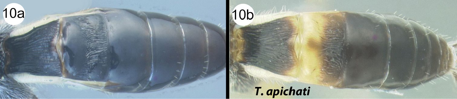
Figure 4MT2 pale in anterior half, melanic posteriorly. Ocellar triangle melanic, concolorous with remainder of vertex.
Body length. 2.6 mm.
Head. Space between antennal insertions with a weakly developed bulge that is weakly declivous posteriorly, dorsal surface of bulge with a shallow longitudinal groove. Number of flagellomeres 23. Posterior surface of scutellum mostly smooth, posterior scutellar depression well-defined by two large pits separated by a short longitudinal ridge.
Mesosoma. Number of pegs on mid tibia = 5. Number of pegs on hind tibia = 9. Sclerite between metasoma and hind coxa narrow, and lacking a high ridge along its length. Length-width of hind femur 0.596/0.24 = 2.5. 2nd submarginal cell reduced to a small dot, petiole longer than cell is high, or large, cell height subequal to petiole length. Hind wing vein Cub emanates from near mid length of apical margin of subbasal cell, Cub short and weak. Point of notauli intersection heavily sculptured over a wide area. Median lobe of mesoscutum not bulging and not sharply declivous anteriorly. Metapleuron with scattered white setae.
Metasoma. MT1 length distinctly longer than apical width. MT1 with narrowly-spaced longitudinal striae, with some microsculpture between striae, and lacking two pairs of distinctly stronger striae (carinae). MT1 distinctly wider apically than basally. Ratio of widest point of MT1 to narrowest point 0.32/0.22 = 1.5. Length-width ratio of MT1 0.44/0.32 = 1.4. MT2 smooth in most of anterior half anteriad transverse groove, longitudinally striate in transverse groove and area posteriad transverse groove, at least medially. Ovipositor much longer than metasoma, about as long as body or longer.
Color. Body mostly melanic, legs mostly pale; body black except as follows: antenna brown, palpi, labrum and other mouthparts yellow, tegula yellow, fore and mid legs entirely yellow, hind coxa mostly black, hind femur mostly brown, paler apically, hind trochanter, tibia, and tarsus mostly yellow, metasomal mediotergite yellow in anterior half or more, anterior metasomal laterotergites and sternites pale yellow. Tegula yellow, contrasting with predominantly black mesoscutum. Ocellar triangle melanic, concolorous with remainder of vertex. Hind tibia mostly pale, melanic apically and with a subbasal melanic band or lateral spot, or mostly pale, melanic apically only. MT2 pale in anterior half, melanic posteriorly.
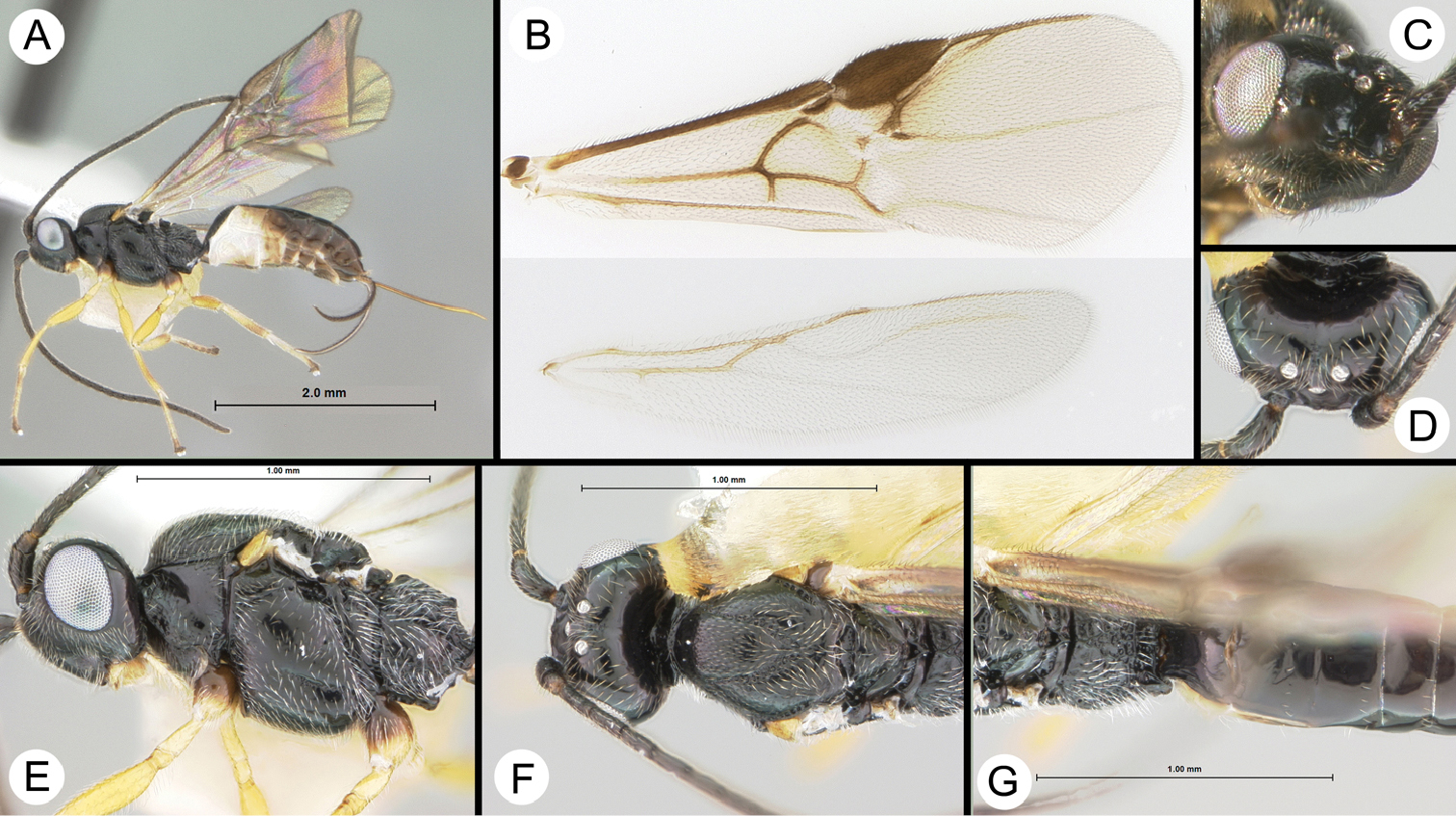
Therophilus apichati sp. n. a lateral habitus b Wings c lateral head and mesosoma d dorsolateral head e dorsal head, mesosoma and propodeum f dorsal Metasoma.
Named in honor of Mr. Apichat Watanawanit, collector for the TIGER project at Doi Chiangdao Wildlife Sanctuary.
H147, GenBank Accession: JQ929183.
Distribution map can be found at http://purl.org/thaimap/apichati
Holotype ♀. H147 [QSBG] Thailand, Khao Kho NP, Mixed deciduous forest, 560m, 16.542°N, 101.042°E, MT, 19–26.xii.2006. http://purl.org/taxabank/T.apichati
urn:lsid:zoobank.org:act:10482AB4-959F-43A7-8E16-88AE03683437
http://species-id.net/wiki/Therophilus_areeluckae
Figure 5MT2 smooth in most of anterior half anteriad transverse groove, longitudinally striate in transverse groove and area posteriad transverse groove, at least medially. Mid femur mostly pale with a bit of melanic color at extreme base. MT2 entirely melanic. Similar to Therophilus rugosiferus but Therophilus areeluckae has no transverse ridge on the propodeum, more sculpture on mesoscutum, and the fore and middle legs are paler.
Body length. 4.0 mm.
Head. Space between antennal insertions with a weakly developed bulge that is weakly declivous posteriorly, dorsal surface of bulge with a shallow longitudinal groove. Number of flagellomeres 28. Posterior surface of scutellum rugose over a semi-circular area that represents the scutellar depression.
Mesosoma. Number of pegs on mid tibia = 5. Number of pegs on hind tibia = 9. Sclerite between metasoma and hind coxa wide with a high ridge along most or all of its length. Length-width of hind femur 0.815/0.256 = 3.2. 2nd submarginal cell reduced to a small dot, petiole longer than cell is high, or large, cell height subequal to petiole length. Hind wing vein Cub emanates from near anterior apex of apical margin of subbasal cell, Cub short and weak. Point of notauli intersection heavily sculptured over a wide area. Median lobe of mesoscutum not bulging and not sharply declivous anteriorly.
Metasoma. MT1 length distinctly longer than apical width. MT1 with narrowly-spaced longitudinal striae, with some microsculpture between striae, and lacking two pairs of distinctly stronger striae (carinae). MT1 distinctly wider apically than basally. Ratio of widest point of MT1 to narrowest point 0.414/0.278 = 1.5. Length-width ratio of MT1 0.668/0.414 = 1.6. MT2 smooth in most of anterior half anteriad transverse groove, longitudinally striate in transverse groove and area posteriad transverse groove, at least medially. Ovipositor much longer than metasoma, about as long as body or longer.
Color. Body mostly melanic, legs mostly pale; body black except as follows: antenna brown, palpi, labrum and other mouthparts yellow; tegula yellow; fore and mid legs yellow except for mostly melanic coxae; hind coxa, trochanter, and femur black; hind tibia mostly pale yellow, melanic apically and with a very weak patch of light brown sub-basally; hind basitarsomere mostly yellow, remaining tarsomeres mostly melanic; anterior metasomal laterotergites and sternites pale yellow; fore wing weakly infuscate. Scape entirely melanic. Tegula yellow, contrasting with predominantly black mesoscutum. Ocellar triangle melanic, concolorous with remainder of vertex. Hind tibia mostly pale, melanic apically and with a subbasal melanic band or lateral spot, or mostly pale, melanic apically only. Fore tarsus mostly or entirely pale. Pronotum entirely melanic. MT2 entirely melanic.
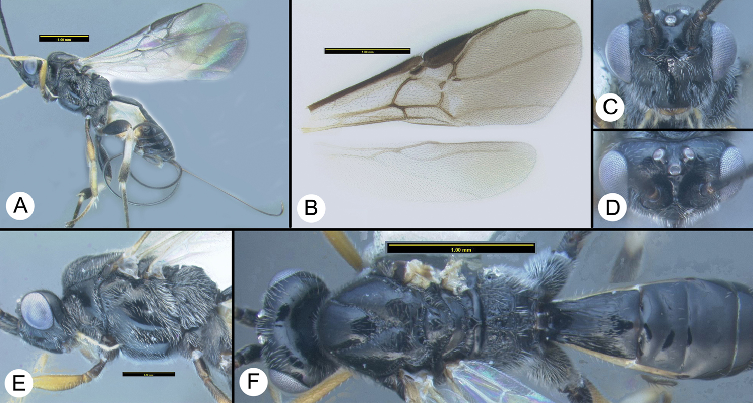
Therophilus areeluckae sp. n. a lateral habitus b Wings c anterodorsal head d dorsal head e lateral head and mesosoma f dorsal head and mesosoma g dorsal propodeum and Metasoma.
Named in honor of Ms. Yuwadee Areeluck, collector for the TIGER project at Doi Inthanon National Park.
Distribution map can be found at http://purl.org/thaimap/areeluckae
Holotype ♀. H988 [QSBG] Thailand, Chae Son NP, Youthcamp/meeting hall, 476m, 18.831°N, 99.47°E, MT, 22–28.iii.2008. http://purl.org/taxabank/T.areeluckae
urn:lsid:zoobank.org:act:EC88E916-3270-4275-8333-362B0535B046
http://species-id.net/wiki/Therophilus_boonthami
Figure 6Ocellar triangle melanic, contrasting with remainder of vertex, which is pale. Hind tibia mostly melanic, pale color restricted to extreme base.
Body length. 3.3 mm.
Head. Space between antennal insertions with a weakly developed bulge that is weakly declivous posteriorly, dorsal surface of bulge with a shallow longitudinal groove. Number of flagellomeres 28. Posterior surface of scutellum posterior scutellar depression represented by two pits.
Mesosoma. Number of pegs on mid tibia = 5. Number of pegs on hind tibia = 9. Sclerite between metasoma and hind coxa narrow, and lacking a high ridge along its length. Length-width of hind femur 0.755/0.31 = 2.4. 2nd submarginal cell large, cell height subequal to petiole length. Hind wing vein Cub emanates from near anterior apex of apical margin of subbasal cell, Cub long and weak. Notauli meeting but sculpture not extending outside of well-defined grooves.
Metasoma. MT1 length only slightly longer than apical width. MT1 with narrowly-spaced longitudinal striae, with some microsculpture between striae, and lacking two pairs of distinctly stronger striae (carinae). Ratio of widest point of MT1 to narrowest point 0.481/0.338 = 1.4. Length-width ratio of MT1 0.55/0.481 = 1.1. MT2 entirely smooth. Ovipositor clearly shorter than body, about as long as Metasoma.
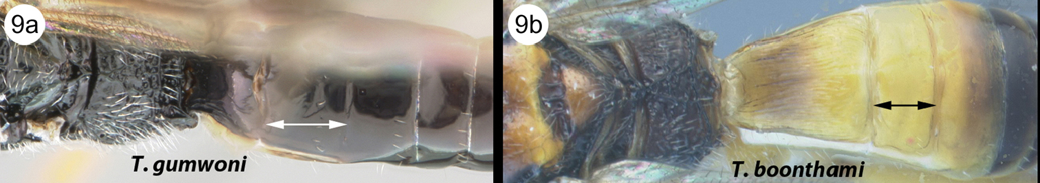
Color. Mostly yellow or yellow-orange with some brown and black; head yellow or orange except antenna, vertex, and occiput brown; thorax yellow or orange and brown, darker near crenulae and sutures; propodeum mostly dark brown; fore and mid legs yellow; hind leg mostly yellow except most of tibia and tarsus melanic, hind tibial spurs yellow; metasoma mostly yellow; posterior terga mostly brown, MT1 with some weak melanic color on longitudinal striae; fore wing weakly infuscate. Tegula yellow, contrasting with predominantly black mesoscutum, or yellow or orange, similar in color to predominantly orange or yellow mesoscutum. Ocellar triangle melanic, contrasting with remainder of vertex. Hind tibia mostly melanic, pale color, if present, restricted to extreme base. MT2 entirely or almost entirely pale.
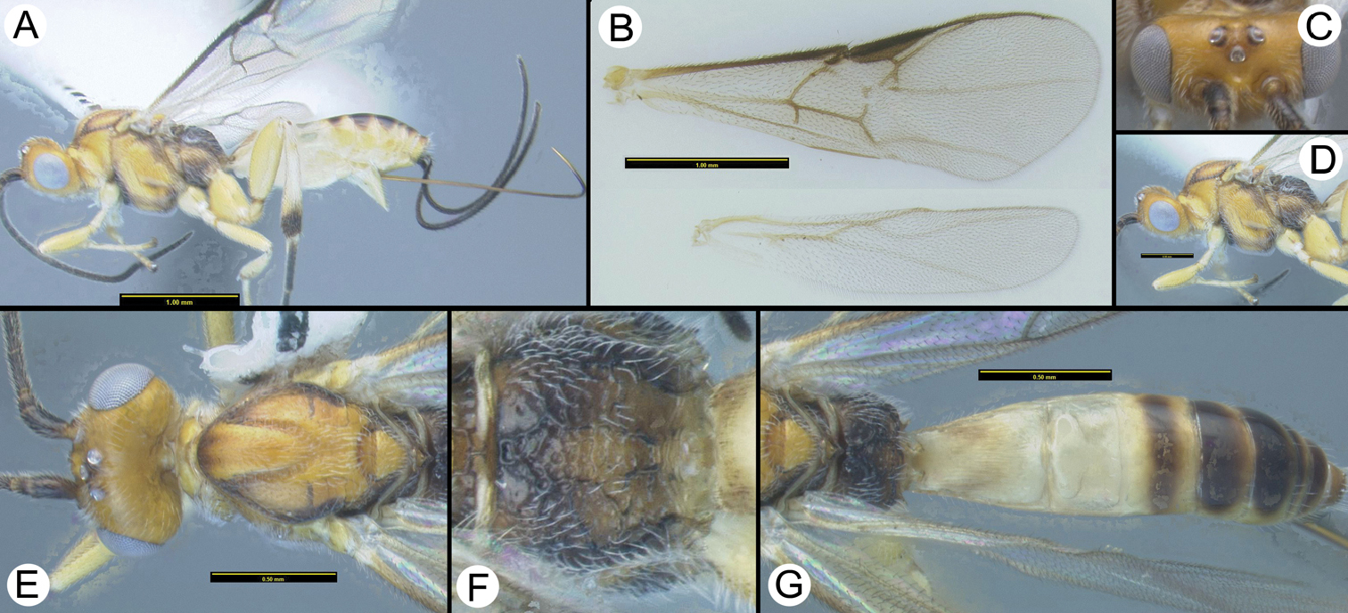
Therophilus boonthami sp. n. a lateral habitus b Wings c anterodorsal head d dorsal head e lateral head and mesosoma f dorsal habitus.
Named in honor of Mr. Tawatchai Boontham, collector for the TIGER project at Huay Namdung National Park.
H633, GenBank Accession: JQ929201.
Distribution map can be found at http://purl.org/thaimap/boonthami
Material examined. Holotype ♀. H633 [QSBG] Thailand, Kaeng Krachan NP, km33/helipad, 735m, 12.836°N, 99.345°E, MT, 18–25.ii.2009. http://purl.org/taxabank/T.boonthami
urn:lsid:zoobank.org:act:14069DD0-B68F-4D92-9058-3C20329DDAA7
http://species-id.net/wiki/Therophilus_chiangmaiensis
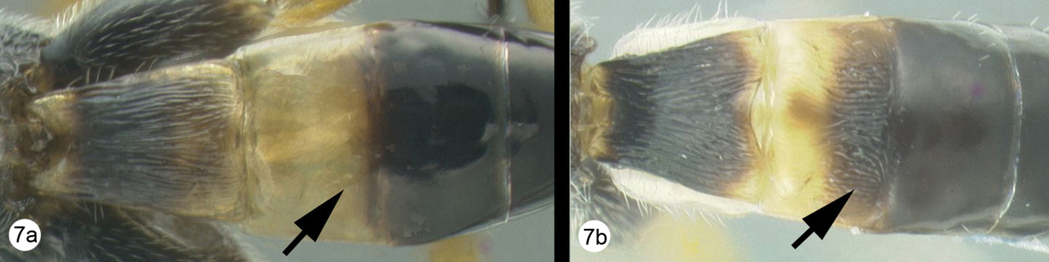
Figure 7Hind tibia mostly melanic with pale coloration restricted to the medial surface at midlength. Wings relatively deeply infuscate.
Body length. 4.6 mm.
Head. Space between antennal insertions with a weakly developed bulge that is weakly declivous posteriorly, dorsal surface of bulge with a shallow longitudinal groove. Number of flagellomeres 28. Posterior surface of scutellum posterior scutellar depression represented by a deep sculptured semicircular pit.
Mesosoma. Number of pegs on mid tibia = 5. Number of pegs on hind tibia = 9. Sclerite between metasoma and hind coxa narrow with a high ridge along most or all of its length. Length-width of hind femur 1.0/0.351 = 2.9. 2nd submarginal cell large, cell height subequal to petiole length. Hind wing vein Cub emanates from near anterior apex of apical margin of subbasal cell, Cub long and weak. Point of notauli intersection heavily sculptured over a wide area with a median longitudinal ridge. Metapleuron with white setae of moderate density.
Metasoma. MT1 with narrowly-spaced, longitudinal striae, with some microsculpture between striae, and with and two pairs of slightly stronger striae (carinae). MT1 distinctly wider apically than basally. Ratio of widest point of MT1 to narrowest point 0.6/0.357 = 1.7. Length-width ratio of MT1 0.77/0.6 = 1.3. MT2 entirely smooth. Ovipositor much longer than metasoma, about as long as body or longer.
Color. Melanic except as follows: mouthparts mostly yellow; mid and hind tibial spurs yellow; hind tibia with a yellow patch medially at mid length; anterior metasomal laterotergites and sternites mostly pale yellow; fore wing infuscate, more so than other species in this revision. Tegula black, concolorous with mesoscutum. Ocellar triangle melanic, concolorous with remainder of vertex. Hind tibia mostly melanic with pale coloration at midlength at least medially. MT2 entirely melanic.
Therophilus chiangmaiensis sp. n. a lateral habitus b Wings c anterodorsal head d dorsal head e lateral head and mesosoma f dorsal head and mesosoma g dorsal propodeum h dorsal Metasoma.
Named after the province in which the type specimen was collected.
H1853, GenBank Accession: JQ929190.
Distribution map can be found at http://purl.org/thaimaps/chiangmaiensis.
Holotype ♀. 1853 [QSBG] Thailand, Chiang Mai, Doi Phahompok NP, Kiewlom1: Montane Forest, 20.0575°N, 99.1425°E, MT 7, 14.viii.2007. http://purl.org/taxabank/T.chiangmaiensis
http://species-id.net/wiki/Therophilus_cattienensis
Figure 8Ocellar triangle pale, concolorous with remainder of vertex. Scape at least partly pale, especially anteriorly.
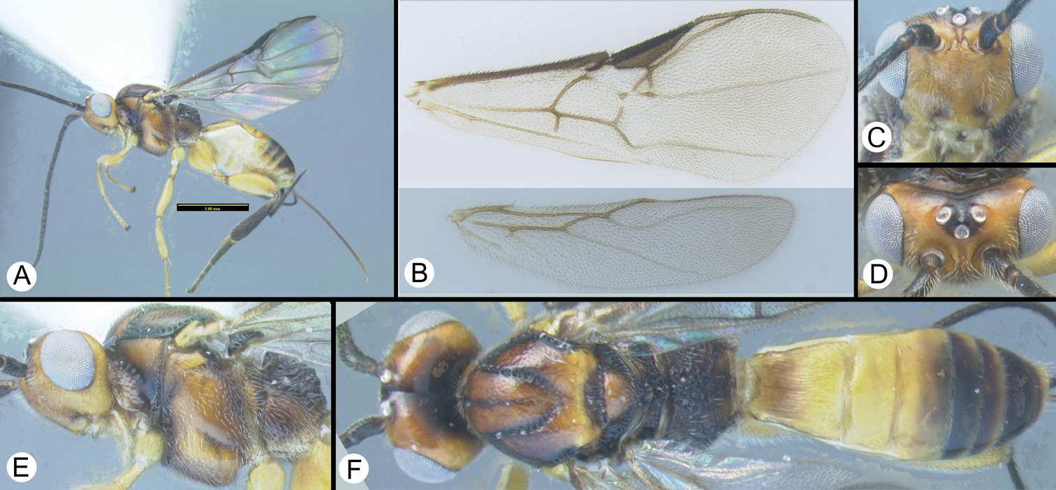
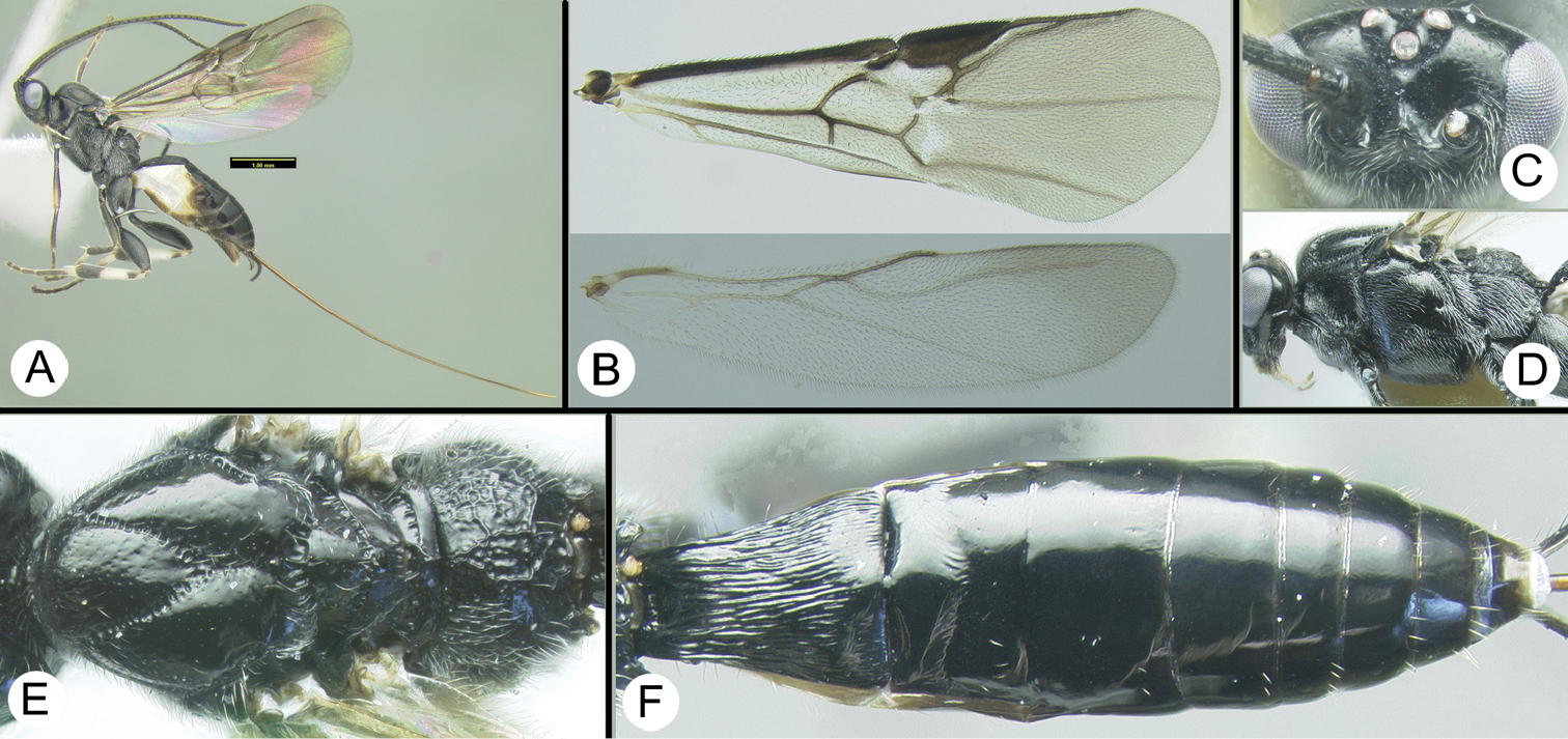
Therophilus cattienensis van Achterberg and Long a lateral habitus b Wings c anterodorsal head d lateral mesosoma e dorsal mesosoma f dorsal propodeum g MT1–MT4.
The Thai specimens differ from the holotype only in the color of the metapleuron which is yellow-brown in the type and melanic in all Thai specimens. This same variation is found in Vietnamese males described by
H024, GenBank Accession: JQ929199; H050, GenBank Accession: JQ929198; H051, GenBank Accession: JQ929197; H059, GenBank Accession: JQ929196; H401, GenBank Accession, JQ929200.
Distribution map can be found at http://purl.org/thaimap/cattienensis
♀. Thailand: Doi Inthanon NP: Vachirathan Fall, 700m, 18.539°N, 98.601°E, MT, 9–16.iii.2007: H0024; 16–23.iii.2007: H0051; 29.iv–6.v.2007: H0058, H0059. H058; Doi Inthanon NP: Kew Maepan Trail, 2200m, 18.553°N, 98.48°E, MT, 29.iv-6.v.2007: H0050. Namtok Mae Surin NP, 19.344°N, 97.988°E, MT, 4–11.v.2008: H0318, H0325, H0329, H0330, H0338, H2427, H2429, H2433, H0471; 19.344°N, 97.988°E, 27.iv–4.v.2008: H0401, H0482; 19.348°N, 97.985°E, 27.iv-4.v.2008: H0482; 18–25.v.2008: H0435; 19.3482°N, 97.9835°E, H3828, H5514. Doi Chiangdao NP, Headquarter, 19.4046°N, 98.9218°E, MT: H5533. Phu Ruea NP, Pah Lo Noy, 1343m, 17.508°N, 101.348°E, MT, 19–26.ix.2006: H5931, H5933. Khao Sok NP, Headquarter, 115m, 8.915°N, 98.53°E, MT, 25.xi–2.xii.2008: H0319. Chiang Mai Province, Pa Huay Kho, 20–30.vi.1997: H1120. Depository: H1120, H051, H3828, H059, H5533, H050, H024, H401, H5514, H325, H319, H338, H329, H318, H330, H058 [QSBG]; H5933, H5931, H482, H2429, H471, H435, H2427, H2433, H5535 [HIC]. http://purl.org/taxabank/T.cattienensis
http://species-id.net/wiki/Therophilus_crenulisulcatus
Figure 9Tegula black, concolorous with mesoscutum. 2nd submarginal cell height subequal to petiole length. Hind tibia mostly pale, melanic apically and with a subbasal melanic band or lateral spot. Pronotum entirely melanic. MT2 with weak short longitudinal striae restricted to transverse depression, or entirely smooth.
Therophilus crenulisulcatus van Achterberg & Long a lateral habitus b Wings c anterodorsal head d lateral head and mesosoma e dorsal head and mesosoma f dorsal propodeum and MT1–3
The Thai specimen has a slightly longer ovipositor, otherwise very similar to type.
Distribution map can be found at http://purl.org/thaimap/crenulisulcatus
Material examined. ♀. 8481 [QSBG] Thailand, Doi Phahompok NP, Kiewlom1: Montane Forest, 20.0575°N, 99.1425°E, MT, 7-14.viii.2007. http://purl.org/taxabank/T.crenulisulcatus
urn:lsid:zoobank.org:act:559CE2A4-FCE2-4EBA-A413-EF983FF0AC1B
http://species-id.net/wiki/Therophilus_kwanuiae
Figure 102nd submarginal cell reduced to a small dot, petiole longer than cell is high. Tegula black, concolorous with mesoscutum. Fore tarsus entirely pale.
Body length. 3.8 mm.
Head. Space between antennal insertions with a weakly developed bulge that is weakly declivous posteriorly, dorsal surface of bulge with a shallow longitudinal groove. Number of flagellomeres 29. Posterior surface of scutellum posterior scutellar depression represented by several (2–3) pits.
Mesosoma. Number of pegs on mid tibia = 5. Number of pegs on hind tibia = 8. Sclerite between metasoma and hind coxa narrow with a high ridge along most or all of its length. Length-width of hind femur 0.911/0.292 = 3.1. 2nd submarginal cell reduced to a small dot, petiole longer than cell is high. Hind wing vein Cub emanates from near anterior apex of apical margin of subbasal cell, Cub short and weak. Notauli meeting but sculpture not extending much outside of well-defined grooves.
Metasoma. MT1 length distinctly longer than apical width. MT1 with narrowly-spaced, longitudinal striae, with some microsculpture between striae, and with and two pairs of slightly stronger striae (carinae). MT1 distinctly wider apically than basally. Ratio of widest point of MT1 to narrowest point 0.475/0.3 = 1.6. Length-width ratio of MT1 0.695/0.475 = 1.5. MT2 entirely smooth. Ovipositor much longer than metasoma, about as long as body or longer.
Color. Mostly melanic except as follows: mouthparts yellow except galea melanic; fore and mid legs yellow except coxae, trochanters and base of femora melanic; hind tibia mostly pale, melanic in apical third with a pale, tan, lateral, spot near base; anterior metasomal laterotergites and sternites mostly pale yellow; fore wing weakly infuscate. Tegula black, concolorous with mesoscutum. Ocellar triangle melanic, concolorous with remainder of vertex. Hind tibia mostly pale, melanic apically and with a subbasal melanic band or lateral spot, or mostly pale, melanic apically only. Fore tarsus mostly or entirely pale. MT2 entirely melanic.
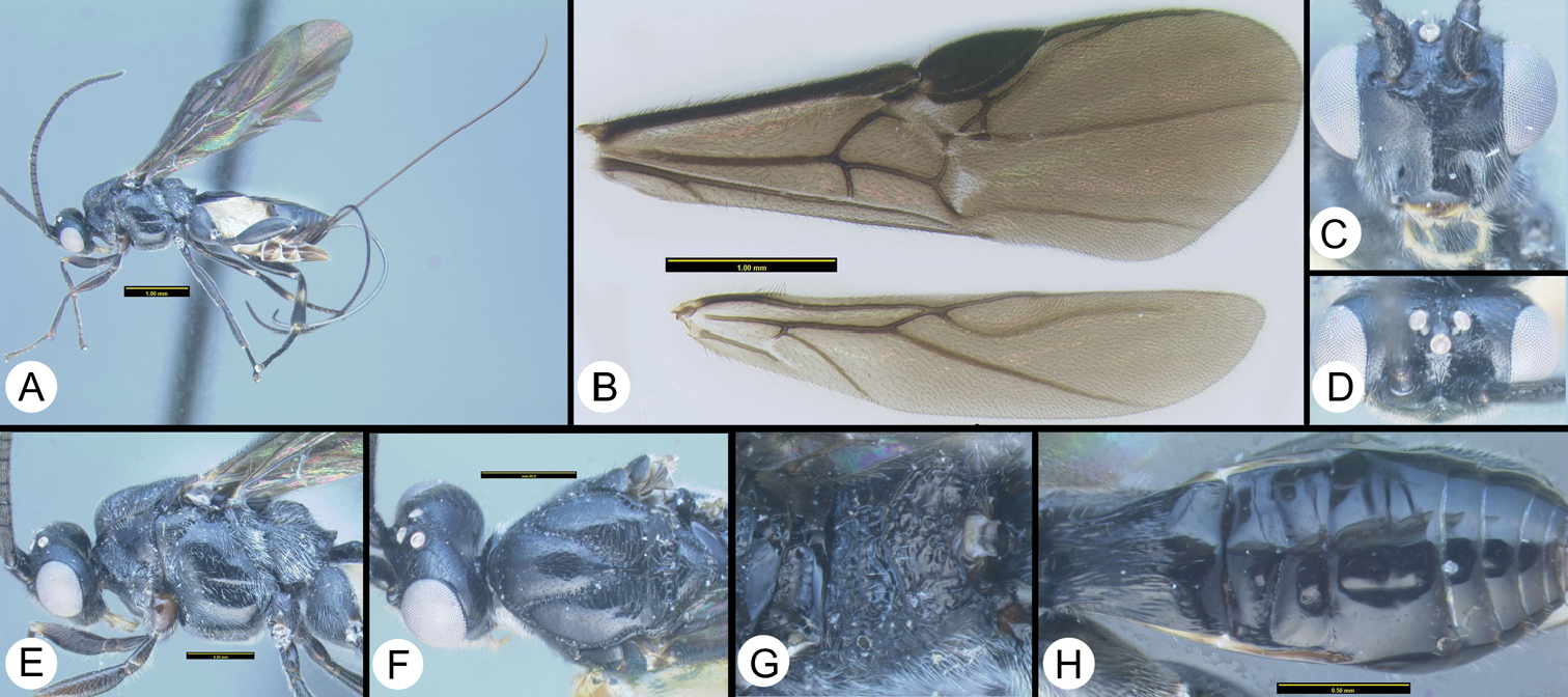
Therophilus kwanuiae n. sp. a lateral habitus b wings c dorsolateral head d dorsal head e lateral head and mesosoma f dorsal head and mesosoma g dorsal propodeum and metasomal terga 1–3.
Named in honor of Ms. Boonruen Kwanui, collector for the TIGER project at Chae Son National Park
Distribution map can be found at http://purl.org/thaimap/kwanuiae
Holotype ♀. H927 [QSBG] Thailand, Huai Nam Dang NP, Visitor center, 19.313°N, 98.607°E, MT, 31.iii–7.iv.2008.
Paratype ♀. H5524 [QSBG] Thailand, Chiang Mai , Huai Nam Dang NP, Thung Buatong View Point , 19.2926°N, 98.6004°E, MT, 7–13.ii.2008 http://purl.org/taxabank/T.kwanuiae
http://species-id.net/wiki/Therophilus_planifrons
Figure 11Ovipositor clearly shorter than body, about as long as Metasoma. Hind tibia mostly pale, melanic apically and with a subbasal melanic band or lateral spot.
Therophilus planifrons van Achterberg & Long. a lateral habitus b Wings c dorsolateral head d dorsal head e lateral head and mesosoma f dorsal head and mesosoma g dorsal propodeum and MT1–MT3.
H235, GenBank Accession: JQ929182.
Distribution. Distribution map can be found at http://purl.org/thaimap/planifrons
♀. H235 [QSBG] Thailand, Huai Nam Dang NP, behind visitor house, 1670m, 19.312°N, 98.607°E, MT, 31.vii–7.viii.2007. http://purl.org/taxabank/T.planifrons
urn:lsid:zoobank.org:act:10BACCAA-3356-4484-B8AA-138F5BA8D516
http://species-id.net/wiki/Therophilus_songrani
Figure 12Ocellar triangle pale, concolorous with remainder of vertex. Tegula yellow or orange, similar in color to predominantly orange or yellow mesoscutum. MT1 distinctly longer than apical width.
Body length. 3.5 mm.
Head. Space between antennal insertions with a weakly developed bulge that is weakly declivous posteriorly, dorsal surface of bulge with a shallow longitudinal groove. Number of flagellomeres 29. Posterior surface of scutellum posterior scutellar depression represented by two pits.
Mesosoma. Number of pegs on mid tibia = 6. Number of pegs on hind tibia = 10. Sclerite between metasoma and hind coxa narrow, and lacking a high ridge along its length. Length-width of hind femur 0.750/0.265 = 2.8. 2nd submarginal cell large, cell height subequal to petiole length. Hind wing vein Cub emanates from near anterior apex of apical margin of subbasal cell, Cub long and weak. Notauli barely meeting and sculpture not extending past meeting point.
Metasoma. MT1 length distinctly longer than apical width. MT1 with narrowly-spaced longitudinal striae, with some microsculpture between striae, and lacking two pairs of distinctly stronger striae (carinae). MT1 distinctly wider apically than basally. Ratio of widest point of MT1 to narrowest point 0.367/0.264 = 1.5. Length-width ratio of MT1 0.54/0.387 = 1.4. MT2 with transverse and/or diagonal striae in and/or near transverse depression. Ovipositor much longer than metasoma, about as long as body or longer.
Color. Yellow or yellow-orange except as follows: antenna melanic; thorax with melanic infusions along crenulae and sutures; propodeum mostly melanic; MT3-MT7 mostly brown; Wings hyaline. Scape entirely melanic. Tegula yellow or orange, similar in color to predominantly orange or yellow mesoscutum. Ocellar triangle pale, concolorous with remainder of vertex. Hind tibia mostly pale, melanic apically only. MT2 entirely or almost entirely pale.
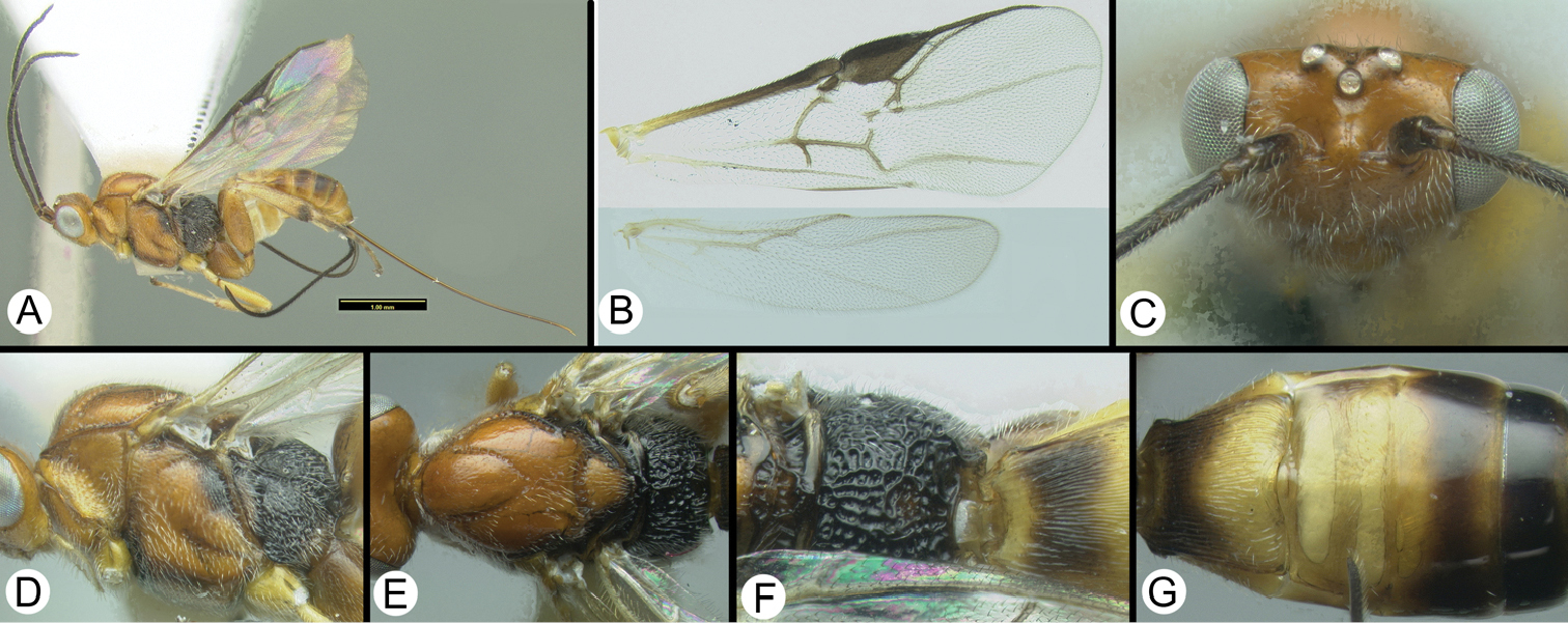
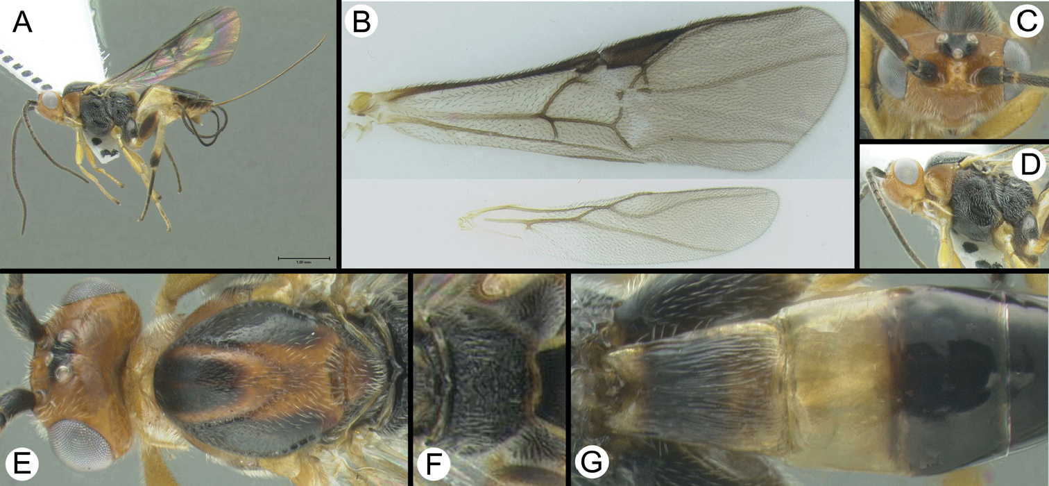
Therophilus songrani sp. n. a lateral habitus b Wings c dorsal head d lateral head and mesosoma e dorsal head and mesosoma f dorsal propodeum g dorsal propodeum and Metasoma.
Named in honor of Mr. Songran Chaksu, collector for the TIGER project at Doi Chiangdao Wildlife Sanctuary.
H352, GenBank Accession: JQ929192.
Distribution map can be found at http://purl.org/thaimap/songrani
Holotype ♀. H352 [QSBG] Thailand, Queen Sirikit Botanic Garden, 811m, 18.881°N, 98.862°E, MT, 30.iv–12.v.2009. http://purl.org/taxabank/T.songrani
urn:lsid:zoobank.org:act:F2A17DCC-B414-44FB-80B8-5B1360C28707
http://species-id.net/wiki/Therophilus_sukpengae
Figure 13MT2 smooth in most of anterior half anteriad transverse groove, longitudinally striate in transverse groove and area posteriad transverse groove, at least medially. Tegula black, concolorous with mesoscutum.
Body length. 3.9 mm.
Head. Space between antennal insertions with a weakly developed bulge that is weakly declivous posteriorly, dorsal surface of bulge with a shallow longitudinal groove. Number of flagellomeres 27. Posterior surface of scutellum posterior scutellar depression represented by two pits.
Mesosoma. Number of pegs on mid tibia = 4. Number of pegs on hind tibia = 9. Sclerite between metasoma and hind coxa narrow with a high ridge along most or all of its length. Length-width of hind femur 0.8/0.266 = 3.0. 2nd submarginal cell large, cell height subequal to petiole length. Hind wing vein Cub emanates from near anterior apex of apical margin of subbasal cell, Cub long and weak. Point of notauli intersection heavily sculptured over a wide area.
Metasoma. MT1 length distinctly longer than apical width. MT1 with narrowly-spaced longitudinal striae, with some microsculpture between striae, and lacking one pair of distinctly stronger striae (carinae). MT1 distinctly wider apically than basally. Ratio of widest point of MT1 to narrowest point 0.383/0.263 = 1.5. Length-width ratio of MT1 0.61/0.383 = 1.6. MT2 smooth in most of anterior half anteriad transverse groove, longitudinally striate in transverse groove and area posteriad transverse groove, at least medially. Ovipositor much longer than metasoma, about as long as body or longer.
Color. Body mostly melanic, legs mostly pale; body melanic except as follows: mouthparts yellow, tegula brown, fore and mid legs mostly yellow except coxae melanic, basal half of femora brown and apex of tibiae and some tarsomeres light brown, hind tibia mostly pale except apex and a light brown subbasal lateral spot, all tibial spurs yellow; anterior metasomal laterotergites and sternites pale yellow; fore wing weakly infuscate. Tegula black, concolorous with mesoscutum. Ocellar triangle melanic, concolorous with remainder of vertex. Hind tibia mostly pale, melanic apically and with a subbasal melanic band or lateral spot. Pronotum entirely melanic. MT2 entirely melanic.

Therophilus sukpengae sp. n. a lateral habitus b Wings c anterior head d lateral head and mesosoma e dorsal head and mesosoma f dorsal propodeum and Metasoma.
Named in honor of Ms. Acharaporn Sukpeng collector for the TIGER project at Chae Son National Park.
Distribution map can be found at http://purl.org/thaimap/sukpengae
Holotype ♀. H998 [QSBG] Thailand Pu Toei NP, Protection unit2/Pu Krathing, 220m, 14.803°N, 99.416°E, MT, 1–7.v.2009. http://purl.org/taxabank/T.sukpengae
urn:lsid:zoobank.org:act:8E959BFE-D614-4100-8123-89A95E0FF0B1
http://species-id.net/wiki/Therophilus_wannai
Figure 14MT2 with short longitudinal striae restricted to transverse depression. Mid femur mostly melanic, pale apically. Fore tarsus mostly pale, melanic basally. Pronotum mostly melanic but with a pale spot dorsomedially.
Body length. 3.8 mm.
Head. Space between antennal insertions with a weakly developed bulge that is weakly declivous posteriorly, dorsal surface of bulge with a shallow longitudinal groove. Number of flagellomeres 28. Posterior surface of scutellum posterior scutellar depression represented by several pits forming a semicircular area.
Mesosoma. Number of pegs on mid tibia = 6. Number of pegs on hind tibia = 8. Sclerite between metasoma and hind coxa narrow with a high ridge along most or all of its length. Length-width of hind femur 0.823/0.256 = 3.2. 2nd submarginal cell large, cell height subequal to petiole length. Hind wing vein Cub emanates from near anterior apex of apical margin of subbasal cell, Cub short and weak. Notauli extending past meeting point but sculpture not extending over a wide area.
Metasoma. MT1 length distinctly longer than apical width. MT1 with narrowly-spaced longitudinal striae, with some microsculpture between striae, and lacking two pairs of distinctly stronger striae (carinae). MT1 distinctly wider apically than basally. Ratio of widest point of MT1 to narrowest point 0.437/0.281 = 1.6. Length-width ratio of MT1 0.702/0.437 = 1.6. MT2 with short longitudinal striae restricted to transverse depression. Ovipositor much longer than metasoma, about as long as body or longer.
Color. Melanic except as follows: mouthparts mostly yellow; fore and mid femora and tibiae yellow-brown, mid leg noticeably darker than fore leg; hind tibia yellow at mid length and basally, melanic apically and subbasally; anterior metasomal laterotergites and sternites pale yellow; fore wing weakly infuscate. Tegula black, concolorous with mesoscutum. Ocellar triangle melanic, concolorous with remainder of vertex. Hind tibia mostly pale, melanic apically and with a subbasal melanic band or lateral spot. Fore tarsus mostly or entirely pale. Pronotum mostly melanic but pale dorsomedially. MT2 entirely melanic.
Therophilus wannai sp. n. a lateral habitus b Wings c anterior head d dorsal head e lateral head and mesosoma f dorsal head and mesosoma g dorsal propodeum h dorsal MT1–MT3.
Named in honor of Mr. Charoen Wanna, collector for the TIGER project at Doi Phuka National Park.
Distribution map can be found at http://purl.org/thaimap/wannai
Holotype ♀. H345 [QSBG] Thailand Doi Phu Kha NP, Office 11, 1359m, 19.208°N, 101.081°E, MT, 15–22.xi.2007. http://purl.org/taxabank/T.wannai
urn:lsid:zoobank.org:act:727FD2FF-CCCD-4090-930D-7DF23FDF71BB
http://species-id.net/wiki/Therophilus_wongchaii
Figure 15Ocellar triangle melanic, contrasting with remainder of vertex, or pale, concolorous with remainder of vertex. Tegula yellow, contrasting with black lateral lobes of mesoscutum. Hind tibia largely pale, melanic apically and with a subbasal melanic band or lateral spot, or mostly pale, melanic apically only.
Body length. 3.5 mm.
Head. Space between antennal insertions with a weakly developed bulge that is weakly declivous posteriorly, dorsal surface of bulge with a shallow longitudinal groove. Number of flagellomeres 29. Posterior surface of scutellum posterior scutellar depression represented by two pits.
Mesosoma. Number of pegs on mid tibia = 5. Number of pegs on hind tibia = 9. Sclerite between metasoma and hind coxa narrow, and lacking a high ridge along its length. Length-width of hind femur 0.850/0.298 = 2.9. 2nd submarginal cell large, cell height subequal to petiole length. Hind wing vein Cub emanates from near anterior apex of apical margin of subbasal cell, Cub long and weak. Notauli extending past meeting point but sculpture not extending over a wide area.
Metasoma. MT1 length distinctly longer than apical width. MT1 with narrowly-spaced longitudinal striae, with some microsculpture between striae, and lacking two pairs of distinctly stronger striae (carinae). MT1 not distinctly wider apically than basally. Ratio of widest point of MT1 to narrowest point 0.4/0.311 = 1.3. Length-width ratio of MT1 0.642/0.4 = 1.6. MT2 with transverse and/or diagonal striae in and/or near transverse depression. Ovipositor much longer than metasoma, about as long as body or longer.
Color. Orange, yellow, black, and brown; head mostly orange, ocellar triangle melanic; antenna melanic; mesoscutum mostly pale medially, melanic laterally; prothorax yellow; meso and metapleuron and propodeum melanic; fore and mid legs yellow; hind leg brown except trochanter and most of tibia yellow; metasomal mediotergites mostly melanic except base of MT1 and all of MT2 yellow; anterior metasomal laterotergites and sternites pale yellow, remainder of metasoma melanic; fore wing weakly infuscate. Scape entirely melanic. Tegula yellow, contrasting with predominantly black mesoscutum. Ocellar triangle melanic, contrasting with remainder of vertex, or pale, concolorous with remainder of vertex. Hind tibia mostly pale, melanic apically and with a subbasal melanic band or lateral spot, or mostly pale, melanic apically only. Fore tarsus mostly or entirely pale. MT2 entirely melanic, or pale in anterior half, melanic posteriorly, or entirely or almost entirely pale.
Therophilus wongchaii sp. n. a lateral habitus b Wings c anterodorsal head d later head and mesosoma e dorsal head and mesosoma f dorsal propodeum g dorsal MT1–MT3.
Named in honor of Mr. Prasit Wongchai, collector for the TIGER project at Doi Phahompok National Park.
H314, GenBank Accession: JQ929194; H977, GenBank Accession: JQ929195; H661, GenBank Accession: JQ929193.
Distribution map can be found at http://purl.org/thaimap/wongchaii
Holotype ♀. H314 [QSBG] Thailand, Kaeng Krachan NP, km33/helipad, 735m, 12.836°N, 99.345°E, MT, 7–14.xi.2008.
Paratypes ♀. Thailand: Kaeng Krachan NP: km33/helipad, 735m, 12.836°N, 99.345°E, MT, 11–18.v.2009: H977; 24.iv–4.v.2009: H661; 17–24.iv.2009: H476; 17–24.iv.2009: H476; 4–11.v.2009: H565, H563; 31.x–7.xi.2008: H604; Pa La-U/waterfall/car park1, 12.536°N, 99.4722°E, pan trap, 2–3.v.2009: H303; 12.536°N, 99.468°E, MT, 4–11.xii.2008: H2439; Panernthung/km30 old lavatory, 970m, 12.825°N, 99.365°E, MT, 11–18.vii.2008: H2404. Depository: H977, H661, H476, H565, H604, [QSBG]; H303, H563, H2439, H2404 [HIC].
urn:lsid:zoobank.org:act:0494CCB9-5FB4-43E1-B807-7E434F43761B
http://species-id.net/wiki/Therophilus_wongwani
Figure 162nd submarginal cell reduced to a small dot, petiole longer than cell is high. Tegula black, concolorous with mesoscutum. Fore tarsus mostly melanic with some pale color apically. Exposed portion of ovipositor distinctly longer than body.
Body length. 4.4 mm.
Head. Space between antennal insertions with a weakly developed bulge that is weakly declivous posteriorly, dorsal surface of bulge with a shallow longitudinal groove. Number of flagellomeres 27. Posterior surface of scutellum completely rugose, posterior scutellar depression not distinct.
Mesosoma. Number of pegs on mid tibia = 8. Number of pegs on hind tibia = 11. Sclerite between metasoma and hind coxa narrow with a high ridge along most or all of its length. Length-width of hind femur 0.854/0.263 = 3.3. 2nd submarginal cell reduced to a small dot, petiole longer than cell is high. Hind wing vein Cub emanates from near mid length of apical margin of subbasal cell, Cub long and strong. Notauli extending past meeting point but sculpture not extending over a wide area.
Metasoma. MT1 length distinctly longer than apical width. MT1 with narrowly-spaced longitudinal striae, with some microsculpture between striae, and lacking two pairs of distinctly stronger striae (carinae). MT1 distinctly wider apically than basally. Ratio of widest point of MT1 to narrowest point 0.533/0.290 = 1.8. Length-width ratio of MT1 0.717/0.533 = 1.4. MT2 with short longitudinal striae restricted to transverse depression. Ovipositor much longer than metasoma, about as long as body or longer. Ovipositor length exposed portion of ovipositor distinctly longer than body.
Color. Black except as follows; mouthparts mostly yellow; hind tibia pale yellow except black at apex and subapically; anterior metasomal laterotergites and sternites pale yellow; fore wing weakly infuscate. Tegula black, concolorous with mesoscutum. Ocellar triangle melanic, concolorous with remainder of vertex. Hind tibia mostly pale, melanic apically and with a subbasal melanic band or lateral spot. Fore tarsus mostly or entirely melanic. MT2 entirely melanic.
Therophilus wongwani sp. n. a lateral habitus b Wings c anterodorsal head d lateral head and mesosoma e dorsal mesosoma and propodeum f dorsal Metasoma.
Named in honor of Mr. Nikom Wongwan, collector for the TIGER project at Doi Phuka National Park.
H028, GenBank Accession: JQ29188; H029, GenBank Accession: HQ929189; H047, GenBank: JQ929186; H048, GenBank Accession: JQ929187; H066, GenBank Accession: JQ929185; H1854, GenBank Accession: JQ929191.
Distribution map can be found at http://purl.org/thaimap/wongwani
Holotype ♀. H028 [QSBG] Thailand, Doi Inthanon NP, Summit marsh, 2500m, 18.589°N, 98.486°E, MT, 23.iii–1.v.2007.
Paratypes ♀. Doi Inthanon NP, Summit marsh, 2500m, 18.589°N, 98.486°E, MT, 8-15.v.2007: H5925, H5926, H5929, H8479, H8480, H066; 23.iii-1.v.2007: H029; 17-24.xi.2006: H048; 22-29.iv.2007: H047; 1-8.xii.2006: H285; 5-12.i.2007: H5934; 2-10.xi.2006: H1854; Doi Chiangdao NP, water reservoir, 549m, 19.407°N, 98.921°E, MT, 18-25.ix.2007: H978, H5510; Doi Phahompok NP, Kiewlom2/Montane Forest 20.0571°N, 99.1425°E: H3803. Depository: H5926, H066, H048, H5510, H285, H5929, H5934 [QSBG]; H029, H5925, H8480, H8479, H978, H1854, H047, H3803 [HIC] http://purl.org/taxabank/T.wongwani
We thank all of the staff at Queen Sirikit Botanic Gardens in Chiang Mai for sorting the many hundreds of samples and for the Thai park staff for running Malaise traps and other collection devices. Thanks to Dr. van Achterberg for the loan of type specimens. A special thanks to Chaweewan Hutacharern for managing the Thai end of the TIGER project. Funding was provided by NSF grants DEB-0542864 and EF-0337220.
DELTA data matrix, images, and other files to the dichotomous key for Therophilus s.s. (Hymenoptera: Braconidae: Agathidinae) from Thailand. doi: 10.3897/JHR.27.2832.app1
DELTA data matrix, images, and other files to species descriptions for Therophilus s.s. (Hymenoptera, Braconidae, Agathidinae) from Thailand. doi: 10.3897/JHR.27.2832.app2
Interactive key, in IntKey format, to Therophilus s.s. (Hymenoptera, Braconidae, Agathidinae) from Thailand. doi: 10.3897/JHR.27.2832.app3
Explanation note: To run the identification key, you will need Windows 95/NT or a later version.
You also need to download Intkey software and reboot your computer, if it is not already installed. The software package, Intkey, can be downloaded from http://delta-intkey.com/www/programs.htm. Once Intkey is installed you need only click on the .ink fi le (below) and the key will open. Click on any character on the left to begin.
More details on how to use Intkey efficiently are found at http://florabase.calm.wa.gov.au/help/keys/intkey_tutorial.pdf
Morphological terms matched to the Hymenoptera Anatomy Ontology. Identifiers (URIs) represent anatomical concepts in HAO version http://purl.obolibrary.org/obo/hao/2011-05-18/hao.owl