


(C) 2012 Donald L. J. Quicke. This is an open access article distributed under the terms of the Creative Commons Attribution License 3.0 (CC-BY), which permits unrestricted use, distribution, and reproduction in any medium, provided the original author and source are credited.
For reference, use of the paginated PDF or printed version of this article is recommended.
A new species, Cassidibracon gracillariae Quicke sp. n. from India, is described and illustrated and differentiated from other Indian species. The three known specimens were each reared from distinctive, exposed, bubble-coated cocoons of the gracillariid moth Stomphastis chalybacma (Meyrick, 1908), which superficially resemble insect egg clusters. This is the first reported host record for the genus Cassidibracon Quicke.
Cocoon, Gracillariidae, Plesiobracon group, parasitoid
The Braconinae is one of the largest and most generically diverse subfamilies of braconid parasitic wasps with 177 genera and 2, 442 species described up until 2005 (
During searching of the braconid accessions collection in the Natural History Museum, London, three 100 years old specimens of a small braconine were found, each mounted above a small leaflet with what superficially looked like a white egg-mass. However, on closer examination, these white masses were found to be the distinctive bubble-like ornamentation of the cocoon of a gracillariid moth. The specimens were labelled as having been reared from Epicephala chalybacma, now treated as Stomphastis chalybacma (Meyrick, 1908) (Lepidoptera: Gracillariidae) which is recorded from India and is widespread in S. Asia.
The wasps were all rather dirty, and too fragile to attempt all but the most superficial of cleaning. Nevertheless, they were in sufficiently good condition to be described. The three possess a combination of characters used to describe a putatively monophyletic group of genera referred to as the Plesiobracon
In the key to the Plesiobracon group genera provided by
http://species-id.net/wiki/Cassidibracon
Figures 1–2Head. Terminal flagellomere strongly acuminate. Scapus small, shorter ventrally than dorsally in lateral aspect, not apicolaterally emarginate. Eyes moderately large, glabrous, not emarginate. Malar suture well-developed. Dorsal margin of hypoclypeal depression not strongly protruding and lamelliform. Clypeus sharply demarkated from face by deep groove. Anterior tentorial pits large. Frons not inpressed behind antennal sockets, with strong midlongitudinal groove. Head strongly contracted behind eyes.
Mesosoma. Mesoscutum shiny, moderately densely with punctures at the bases of setae. Notauli very weak anteriorly, not impressed on dorsal surface. Mesopleuron smooth and shiny, largely glabrous. Precoxal suture not impressed. Pleural suture finely crenulate. Median area of metanotum with complete midlongitudinal carina. Midlongitudinal propodeal carina complete. Lateral carinae of propodeum absent, propodeum and metapleuron separated by deep groove.
Wings. Wings evenly setose. Forewing vein 1-SR+M virtually straight. Veins 1-SR and C+SC+R forming angle of aproximately 70°. 2nd submarginal cell trapezoidal. Fore wing vein 1r-m with 2 distinct bullae. Hind wing vein 1-M more than 6 × length or r-m. Hind wing vein 2-SC+R longitudinal.
Legs. Hind tibia robust, without disting longitudinal groove. Claws with small, acutely pointed basal lobes.
Metasoma. Metasoma short and robust, with 5 exposed, coarsely sculptured tergites. 1st tergite with spiracle approximately at midlength, with complete though somewhat irregular dorso-lateral carina behind spiracle, with dorsal carinae fused to form a semicircular transverse carina that runs far closer to posterior margin of tergite than its base. 2nd+3rd tergites large, their combined medial length more than 3 × length of exposed (coarsely sculptured) parts of tergites 4 and 5. 2nd tergite without midbasal or anterolateral areas, with weak sublateral, posteriorly converging grooves on anterior 0.5 2nd suture curved, narrow, crenulate. 3rd tergite without anterolateral areas. Tergites without transverse subposterior grooves. Ovipositor short, sheaths approximately 1.2 × length of hind basitarsus.
Cassidibracon
The type specimens of the three oriental species described by
urn:lsid:zoobank.org:act:A7AE1BB5-7C5C-44F8-8606-6E215FF89DCC
Holotype. Female, “19.vi.1911, Parasite on Epicephala chalybacma, Pusa [INDIA], C. S. Misra”, “72” (BMNH)
Paratypes. 2 females, same data as holotype.
In
| 1 | Propodeum with complete midlongitudinal carina. Body yellowish brown | 2a |
| – | Propodeum with incomplete midlongitudinal carina. Body brown or blackish brown | 3 |
| 2a | Metasoma entirely yellowish. Face with midlongitudinal ridge which is produced to form knob between antennal sockets [Afrotropical] | Cassidibracon castus |
| – | Metasoma with distinct pattern of dark marks. Face without midlongitudinal ridge [Oriental] | 2b |
| 2b | Antenna with 21 flagellomeres. Dark posterior marking on tergite 2 and anterior of tergite 4 entire | Cassidibracon sumodani |
| – | Antenna with 24 flagellomeres. Dark posterior marking on tergite 2 and anterior of tergite 4 completely divided medially by pale brown yellow zone giving rise to ‘H’-shaped pattern | Cassidibracon gracillariae sp. n. |
Length of body 2.9 mm, of forewing 2.6 mm and of antenna 2.8 mm.
Head. Antenna with 24 flagellomeres. Median flagellomeres approximately 1.4 × longer than wide. 1st flagellomere 1.1 × longer than both the 2nd and 3rd segments separately. Face shiny with numerous punctures at bases of setae. Height of eye: width of head: width of face = 1.0: 2.5 : 1.05. Intertentorial distance 1.7 × tentorio-ocular distance. POL: transverse diameter of posterior ocellus: shortest distance between posterior ocellus and eye = 1: 1 : 3.
Mesosoma. Mesosoma approximately 1.5 × longer than high. Midlongitudinal propodeal carina running within a deep, foveolate groove. Propodeum largely shiny, anteriorly smooth becoming distinctly weakly longitudinally striate medially merging to punctate sculpture posteriorly.
Wings. Fore wing vein cu-a marginally postfurcal. Lengths of fore wing veins r:3-SR:SR1 = 1.0: 1.7: 5.0.
Legs. Length of hind femur: tibia: tarsus = 1.2: 1.0: 1.0. Hind tibia 4.5 × longer than maximally deep.
Metasoma. Metasomal tergites irregularly densely punctulate. 2nd tergite 1.9 × wider than medially long. 3rd tergite 2.5 × wider than medially long.
Coloration. Antenna orange-brown becoming black on apical third. Head cream-yellow with stemmaticum black. Mesosoma largely cream-yellow with dark marks on lateral lobes and anterior of middle lobe of mesoscutum. Metasoma cream-coloured with large ‘H’-shaped black mark extending over tergites 2-4.
The type series of Cassidibracon gracillariae sp. n. are labelled as having been reared from ‘E[picephala]. chalybacma’ (now Stomphastis chalybacma (Meyrick, 1908)) (Lepidoptera: Gracillariidae). Specimens of Stomphastis chalybacma in BMNH share the highly distinctive cocoons and there is no doubt that the original host identification was correct. The host is a widespread moth in south-east Asia which mines leaves of Caesalpinia and Samanea species (Fabaceae). The gracillariid, which feeds solitarily as a leaf-miner, pupates in a flattened silken cocoon ornamented with a cluster of distinctive bubbles, excreted by the larva, along the whole length of the cocoon. These bubbles presumably serve a defensive (or camouflage) function and resemble an egg mass, or possibly a parasitoid cocoon mass. The ovipositing Cassidibracon presumably attacks either pre-pupal larva or pupal hosts.
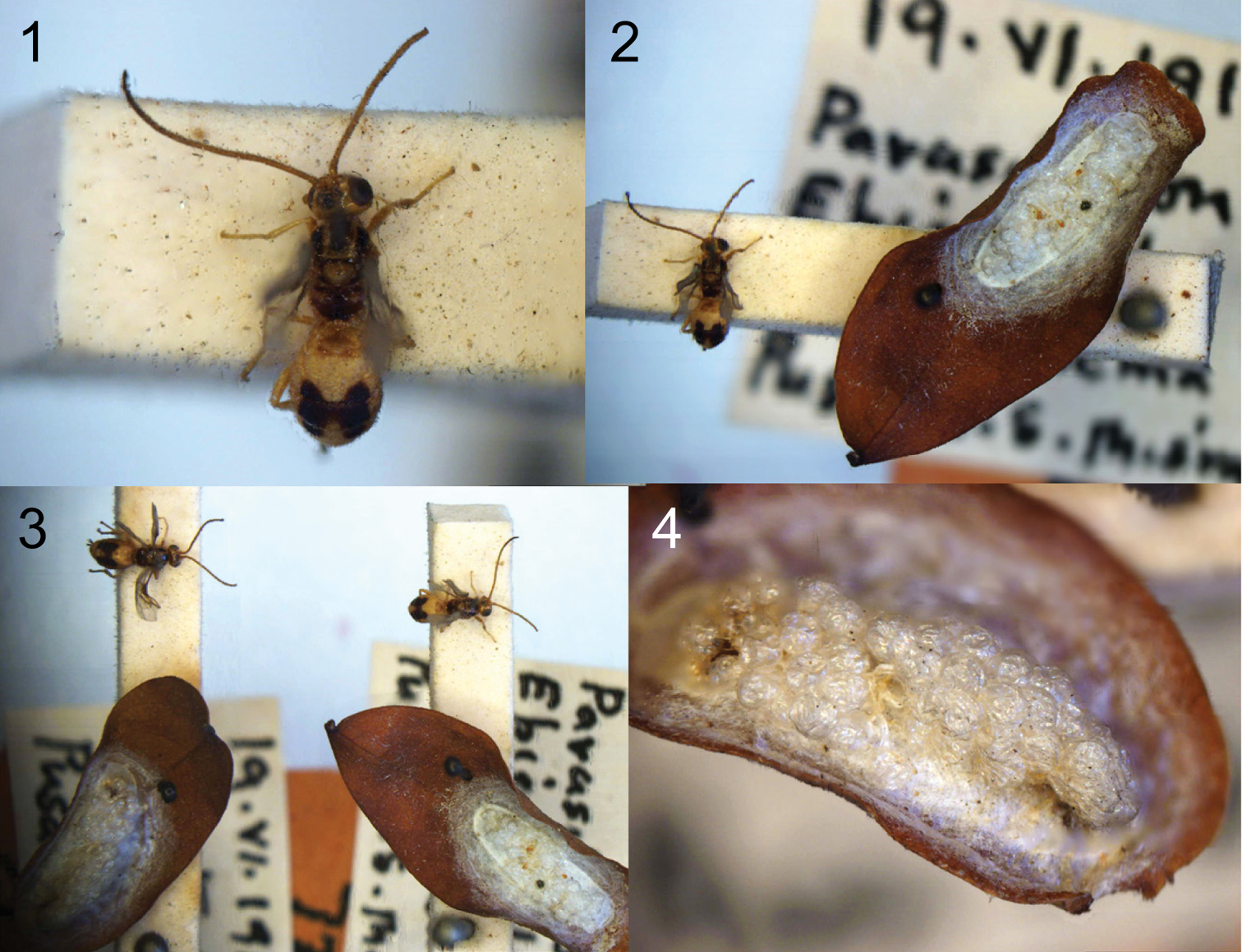
Cassidibracon gracillariae Quicke sp. n., specimens and host cocoon Cell^D® light micrographs. 1 holotype, habitus 2 holotype and associated host remains 3 holotype and paratype 4 detail of host remains of paratype.
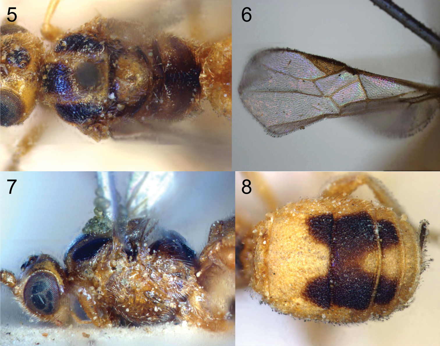
Cassidibracon gracillariae Quicke sp. n., holotype, Cell^D® light micrographs. 5 posterior of head and mesosoma, dorsal aspect. 6 Fore wing 7 Head and mesosoma, near lateral aspect 8 Metasoma, dorsal aspect.
We are grateful to David Lees, David Notton and Martin Honey (all BMNH) for (independently) confirming the host identity and stage. The Animal Systematic Research Unit and Integrated Ecology Lab, Department of Biology, Faculty of Science, Chulalongkorn University kindly allowed use of their Cell^D® imaging facility.