


(C) 2013 Elijah J. Talamas. This is an open access article distributed under the terms of the Creative Commons Attribution License 3.0 (CC-BY), which permits unrestricted use, distribution, and reproduction in any medium, provided the original author and source are credited.
For reference, use of the paginated PDF or printed version of this article is recommended.
Citation: Talamas EJ, Masner L, Johnson NF (2013) Systematics of Trichoteleia Kieffer and Paridris Kieffer (Hymenoptera, Platygastroidea, Platygastridae). Journal of Hymenoptera Research 34: 1–79. doi: 10.3897/JHR.34.4714
Paridris Kieffer and Trichoteleia Kieffer are morphologically similar genera of solitary egg parasitoids with little overlap between their distributions: Paridris is found commonly worldwide with the exceptions of Madagascar, from which a single specimen is known, and New Zealand, from which no records are known; Trichoteleia is endemic to the Malagasy islands. Here we present the first phylogenetic analysis of platygastroid wasps that combines and compares morphological and molecular data. We find the results of the phylogenetic analyses of the two data sources to be largely congruent for the species treated here. Paridris and Trichoteleia are found to be monophyletic, as are two morphologically well-defined species groups within Paridris. Neoparidris Galloway is found to belong within Paridris and is treated as a junior synonym, syn. n. The faunas of Paridris from Africa, Melanesia and the Indo-Malay islands are revised. Fifteen species are described of which 9 are new: Paridris anikulapo Talamas, sp. n. (sub-Saharan Africa); Paridris densiclava (Kieffer), (Seychelles); Paridris bispinosa (Masner), (Gabon); Paridris nigriclava (Kieffer), (Seychelles); Paridris nitidiceps (Kieffer), (Seychelles); Paridris tenuis (Nixon), (sub-Saharan Africa); Paridris trispinosa Talamas & Masner, sp. n., (Cameroon, Democratic Republic of the Congo); Paridris bifurcata (Dodd), comb. n., (Australia, Papua New Guinea); Paridris mnestros Talamas & Masner, sp. n., (Indonesia, Malaysia); Paridris pantex Talamas, sp. n., (Fiji); Paridris phrikos Talamas & Masner, sp. n., (Fiji); Paridris skolops Talamas & Masner, sp. n., (Fiji); Paridris sulcata Talamas, sp. n., (Vanuatu); Paridris taekuli Talamas & Masner, sp. n., (Australia, Bangladesh, Fiji, India, Indonesia, Ivory Coast, Madagascar, New Caledonia, Thailand, Vietnam); Paridris xestos Talamas & Masner, sp. n., (Fiji). Paridris flaviclava (Kieffer), syn. n., and Paridris nigraticeps (Kieffer), syn. n., are treated as junior synonyms of Paridris nigriclava.
Egg-parasitoid, Platygastroidea, key, Paridris, Trichoteleia, revision, phylogeny
Paridris is a genus of minute wasps that, extrapolating from a single host record from North America (
Among platygastroid genera, the diversity of species revealed by recent taxonomy is often an order of magnitude greater than was previously known (e.g.,
Our focus for this revision is on the geographical regions of Africa, Melanesia and the Indo-Malay islands based on the accessibility of primary types.
Although we were unable to access the type material for the Indian species, the high quality images of Paridris spinosa Rajmohana and the key to Indian species (
Previous phylogenetic analyses of platygastroids have used morphological data (
This work is conducted as part of the Platygastroidea Planetary Biodiversity Inventory and represents a step toward a species-level revision of the Scelionini sensu lato. The contributions of the authors are as follows: E.J. Talamas: DNA extraction and amplification; sequence alignment and phylogenetic analysis, character definition and coding, species concept development, imaging, key development, manuscript preparation; L. Masner: aggregation of specimens, species concept development, manuscript preparation; N.F. Johnson: software and database development, character definition; manuscript preparation.
Primary types: The primary types of J. J. Kieffer and G. E. J. Nixon in The Natural History Museum were photographed by E. Talamas during a visit to this collection in 2009. Our assessment of Neoparidris, and ultimately its treatment as a junior synonym of Paridris, was facilitated by images of the type species taken by N. F. Johnson in 2004 at the Queensland Museum, Brisbane, Australia. Continual access to images of the type material made this project possible without risking damage to specimens during shipping. We hope that this demonstration of the utility of such photographs will encourage the imaging of type material as standard practice in taxonomy.
Specimens: This work is based upon specimens deposited in the following collections, with abbreviations used in the text:
AEIC American Entomological Institute, Gainsville, USA
BMNH Natural History Museum, London, England
BPBM Bishop Museum, Honolulu, USA
CASC California Academy of Sciences, San Francisco, USA
CNCI Canadian National Collection of Insects, Ottawa, Canada
FNIC Fiji National Insect Collection, Suva, Fiji
MCZ Harvard University Museum of Comparative Zoology, Cambridge, USA
MZLU Lund Museum of Zoology, Lund University, Lund, Sweden
MNHN Muséum National d’Histoire Naturelle, Paris, France
OSUC C.A. Triplehorn Insect Collection, Columbus, USA
QSBG Queen Sirikit Botanic Garden, Chiang Mai, Thailand
RMNH Leiden Nationaal Natuurhistorische Museum, Netherlands
SAMC Iziko Museums of Cape Town, South Africa
USNM Smithsonian National Museum of Natural History, Washington DC, USA
Morphological terminology: Abbreviations and morphological terms used in text: A1, A2, ... A12: antennomere 1, 2, ... 12; claval formula: distribution of the multiporous basiconic sensilla on the underside of apical antennomeres of the female, with the antennomere interval specified followed by the number of sensilla per segment (
anterior propodeal projection (app: Figs 12, 75, 79, 92)
felt field (ff: Figs 18–23)
occipital carina (occ: Figs 6, 7, 9)
post gena (pg: Figs 7, 8, 9)
posterior mesepimeral area (pmma: Figs 28, 45)
transverse carina of T2 (trc: Fig. 79).
Morphological terms used in this revision were matched to the Hymenoptera Anatomy Ontology (HAO,
The description of surface sculpture is presented in two formats. Areas of the exoskeleton in which the sculptural elements are inseparable are described simply as “sculpture”. For areas in which the sculptural elements vary independently, sculpture is divided into three categories: punctation: round depressions associated with setae; macrosculpture: raised or sunken patterns of texture that are oriented linearly or radially with respect to punctation or the axes of the body; microsculpture: unoriented, very fine wrinkles or pustulations that occur on, in, or between elements of macrosculpture and punctation.
Information management: The locality data reported for primary types are not a literal transcription of the labels: some abbreviations are expanded; additional data from the collectors are also included. The holotypes should be unambiguously identifiable by means of the unique identifier or the red holotype label. The numbers prefixed with “OSUC ” and “CASENT ” are unique identifiers for the individual specimens (note the blank space after the acronyms). Details on the data associated with these specimens may be accessed at the following link, purl.oclc.org/NET/hymenoptera/hol, and entering the identifier in the form. This monograph also features simultaneous publication and distribution of taxonomic and occurrence records through the Global Biodiversity Information Facility (GBIF) using DarwinCore Archives. All new species have been prospectively registered with Zoobank (
Cybertools: The species descriptions are generated by a database application, vSysLab (purl.oclc.org/NET/hymenoptera/vSysLab), designed to facilitate the generation of taxon by character data matrices, to integrate these with the existing taxonomic and specimen-level database, and to export the data both as text and as input files for other applications (
Imaging: Images were produced using Combine ZP and AutoMontage extended-focus software. The individual images are archived at the image database at The Ohio State University (purl.oclc.org/NET/hymenoptera/specimage) and with MorphBank (www.morphbank.net). The latter also contains collections of images organized by plate.
Species concept: For the purpose of this revision, species are defined as taxa diagnosable by putative autapomorphies or a unique combination of fixed character states.
Molecular data: DNA was extracted nondestructively with a Qiagen DNeasy extraction kit and amplified according to standard protocols with the primers of
Phylogenetic analysis: We analyzed our data under the criterion of parsimony using TNT (
Composite terminals: The CO1 sequence for our outgroup terminal, Archaeoteleia, was amplified from Archaeoteleia mellea, and 18S and 28S sequences from Archaeoteleia onamata. Morphology was coded from Archaeoteleia mellea. For the morphological characters used in our analysis, these species of Archaeoteleia are essentially isomorphic. The sequence data for Paridris aeneus came from two specimens: OSUC 261872 for CO1 and OSUC 265221 for 18S and 28S.
Excluded species: We excluded Paridris armata Talamas, Paridris invicta Talamas, Paridris nitidiceps and Paridris densiclava from our final analyses. Paridris armata, Paridris invicta, and Paridris nitidiceps are known only from males, lacking phylogenetically important female characters, and are of uncertain affinity. The morphological characters of Paridris nitidiceps and Paridris densiclava were coded from photographs of the type specimens. Consequently, we were unable to observe a number of the characters that we consider to be phylogenetically informative. Apart from a loss of resolution, analyses that included these species did not differ from those presented in Figs 2–3.
All of our analyses confirmed monophyly of the Paridris nephta and Paridris pallipes species groups and Trichoteleia (Figs 1–3) with strong bootstrap support for these clades in the molecular and combined analyses. Trichoteleia and the Paridris nephta group had significant support in the morphological analysis, but support for the Paridris pallipes group here was poor, reflecting the homoplasious nature of the characters that delimit this group. Similarly, Paridris was retrieved as a monophyletic group in all of the analyses, but its highest bootstrap value of 49, retrieved in the analysis of combined data, is still low. From a morphological perspective this is unsurprising because all of the synapomorphies for Paridris are also found in other genera or are lost secondarily. The presence of Probaryconus sp. 2 among Paridris in the morphological analysis (Fig. 2) is also not unexpected, particularly because this specimen was selected for its similarity to Paridris: it lacks an epomial carina and has setose compound eyes. Topologically, the only character that separates this species from Paridris is the externally undifferentiated metascutellum. Even this character must be carefully assessed because in some species of both Paridris and Probaryconus the horn of T1 may be very large and preclude observation of the metanotum. However, the high bootstrap support for Probaryconus in the molecular and combined analyses ultimately affirms confidence in our concepts for these genera. A subset of characters that are diagnostic for genera and species treated in this analysis (see Appendix II) are mapped onto the combined data phylogeny in Figure 3.
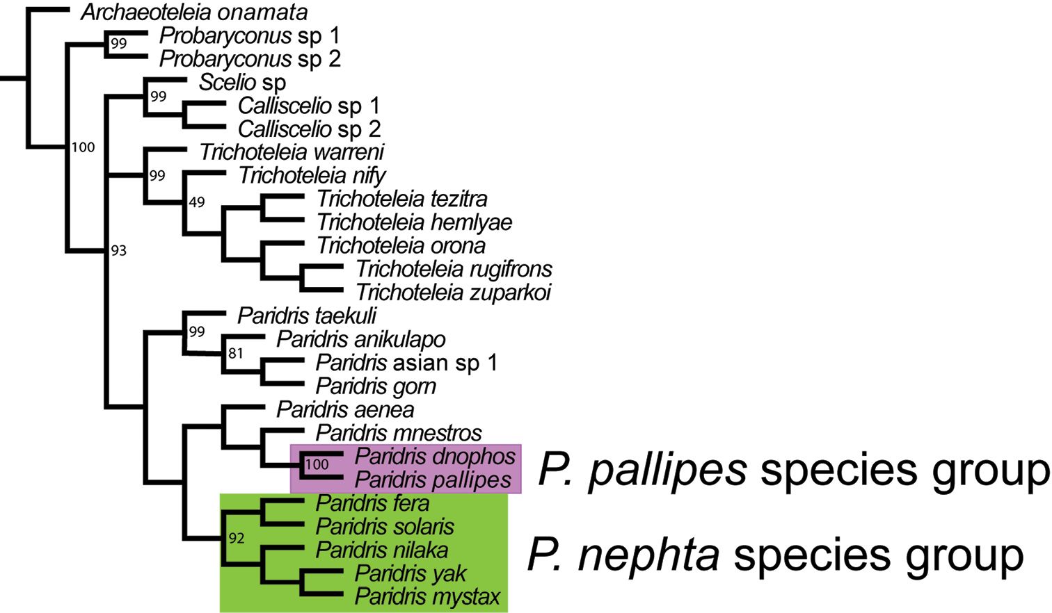
Strict consensus tree based on 18S, 28S and CO1 sequences. Bootstrap support of 49 and higher indicated on tree. CI: 0.515. RI: 0.476.
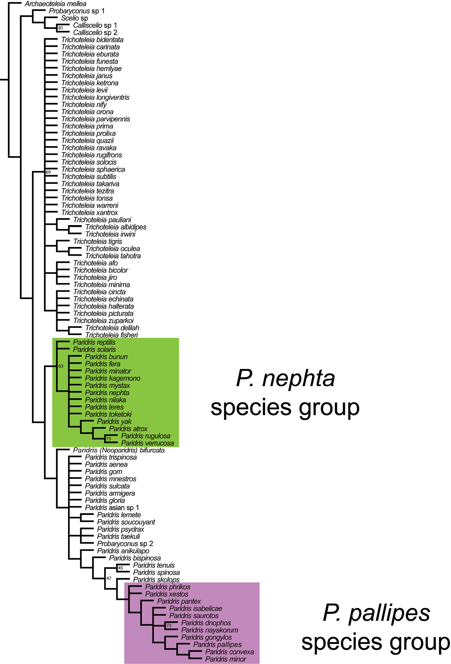
Strict consensus tree based on 72 parsimony informative morphological characters. Bootstrap support of 45 and higher indicated on tree. CI: 0.289. RI: 0.782.
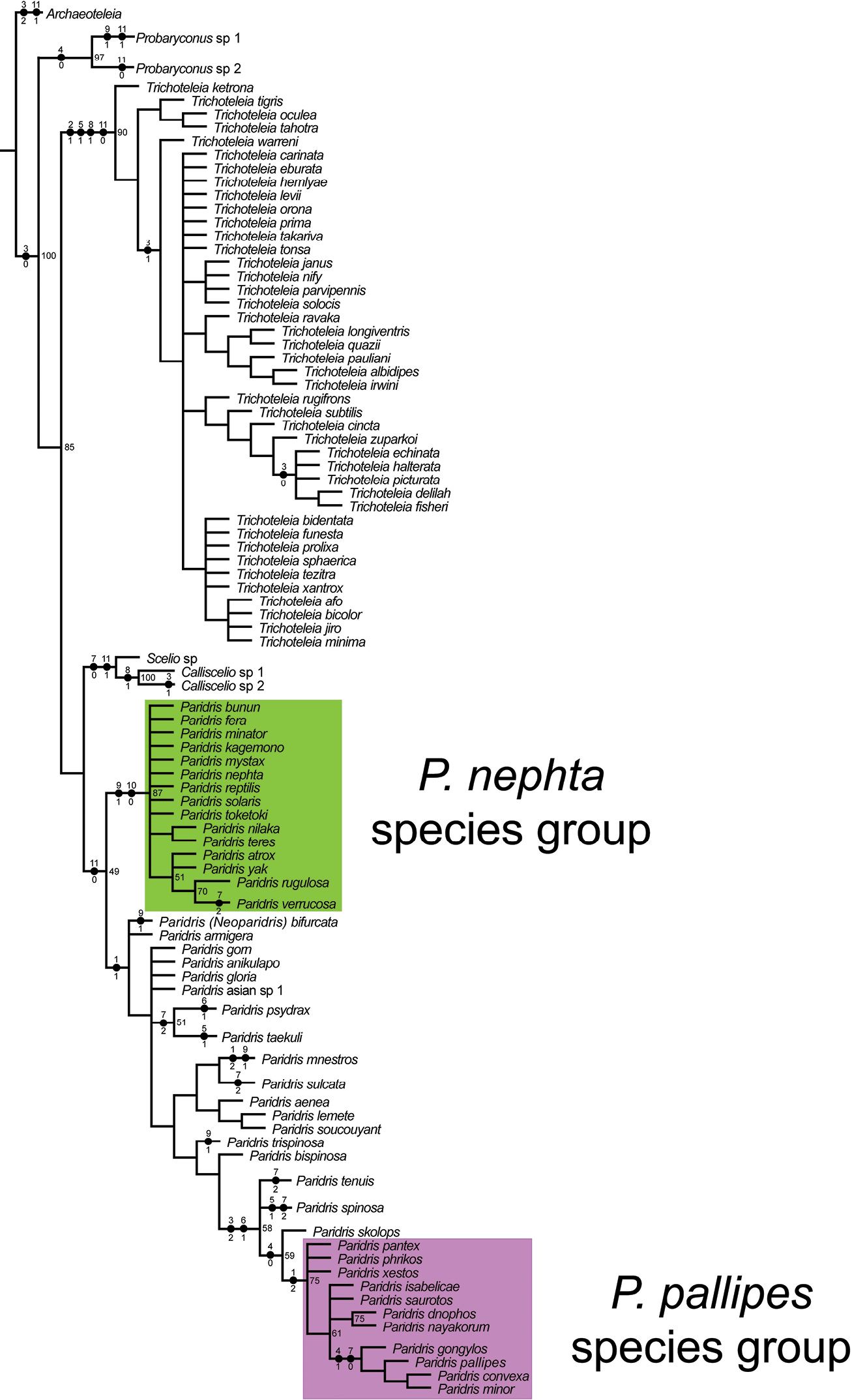
Strict consensus tree based on combined dataset of 72 morphological characters and 18S, 28S, and CO1 sequences. Bootstrap support of 49 and higher indicated on tree. CI: 0.481. RI: 0.585. Black circles indicate the optimization of characters from Appendix II. Numbers above the circles indicate the character; numbers below the circles indicate the character state.
The analysis of Partitioned Bremer Support (see Appendix III) indicates that contributions to clade support from the four data partitions, morphology, 28S, 18S, and CO1, are clade dependent. Within Trichoteleia, 28S provided minimal clade support with values of zero for most of the nodes. CO1 contained the most contrarian signal for this genus and accounted for nearly all of the disagreement between partitions with negative values for six of the twenty nodes. However, none of these were less than negative one, indicating that the incongruence of CO1 with the other data partition is small in magnitude for Trichoteleia. The Paridris nephta species group yielded a similar pattern with no contribution to node support from 28S and a small degree of incongruence from CO1 and 18S. At each node within this group morphology provided the strongest signal.
The pattern of clade support from 18S was consistent with a relatively slow rate of evolution in this gene; nodes at the base of Paridris had 18S support values an order of magnitude higher than those toward the tips. Within the the Scelio+Calliscelio clade the support values for all three genes were the largest (both positive and negative) with the signal of 18S and CO1 conflicting with, and overriding, that of 28S.
Morphological homoplasy within Platygastroidea has been mentioned by previous authors (
We note that the genera within Scelioninae are typically well-defined groups, but are based on unique combinations of characters found throughout the subfamily, that are thus homoplasious in phylogenetic analyses. In polytypic groups, such as Paridris, even the characters that define the group are either secondarily lost (transverse carina on T2), or are apparently homoplasious (metascutellum). Trichoteleia is a group well defined by multiple synapomorphies (felt fields of T1 and S2, setose metascutellum), but these characters do little to ally it with other genera. For example, setation of the eyes is common, and may be present or absent within genera (Idris, Probaryconus). Metascutellar setation, though uncommon, is found in Paridris spinosa and Paridris taekuli as well as in genera that are morphologically more distant (Chromoteleia, Bracalba, Sceliacanthella (OSUC 150176)) and in a species of Teleasinae (OSUC 281605).
The outgroups in our analysis represent a small fraction of the subfamily Scelioninae, and we do not conclude from these results any relationships at the level of genera, only that Trichoteleia is not derived from within Paridris, answering our primary question.
The Paridris pallipes species group is morphologically distinct from the remainder of Paridris, noticeable immediately by the relative absence of macrosculpture from the head and mesosoma. The geographical distribution of this group is perplexing - it is found throughout North and South America and in the Fijian Islands. This suggested the possibility of “tramp” species, yet no species are shared between the two regions. Additionally, Paridris pantex (Fiji) has a highly apomorphic form of the felt fields on S2 that we consider unlikely to have evolved during recent history in which humans have been able to travel rapidly between Fiji and the Americas. It is possible that the group was once widespread, and the distribution we see now is the result of extinction, or that one of the centers of diversity is simply a radiation of the other. Either way, our understanding of the group, and Paridris as a whole, will be greatly furthered by additional host and biological data that allows us to make more informed inferences.
The association between the Paridris pallipes species group and islands is noteworthy. In addition to the three species known from the Fijian islands, 6 of the 8 species of the Paridris pallipes group in the New World are found on Caribbean islands, 4 of them exclusively so.
Diagnosis. The Paridris pallipes species group can be separated from the remainder of Paridris by the following combination of characters: genal striae strongly reduced, rarely extending to midpoint of compound eye; occipital carina absent below foramen magnum; occipital carina complete dorsally; dorsal frons and vertex without macrosculpture; plical carina absent; posterior margin of metascutellum straight to convex; antecostal sulcus of T2 present as a constriction or line of foveae, without carina along its posterior margin; postmarginal vein punctiform.
In addition to these ubiquitously present characters, species of the Paridris pallipes group often have dense setation on the postgena and S1. All species except for Paridris pantex have the felt field present as a line of dense setae along a longitudinal ridge. In a few specimens of Paridris dnophos and Paridris pallipes the lateral ocellus is less than two ocellar diameters from the inner orbit of the compound eye. However, in the vast majority of specimens of these species, and in all other members of this species group, the lateral ocellus is distinctly remote from the inner orbits.
The fauna of Paridris in continental Africa is surprisingly small with just five species. Two of these, Paridris tenuis and Paridris anikulapo, are widespread in distribution and found in eastern, western and southern Africa. Our knowledge about their presence in central Africa, and the existence of other species of Paridris, is currently limited by a dearth of collecting in this region.
Three valid species are known from the Seychelles. Paridris densiclava and Paridris nitidiceps were described by J. J. Kieffer from singletons of opposite sex. We have no additional material of either species, and because we found characters to separate them we consider it best to keep them as separate species. However, we acknowledge that we are currently unable to assess intraspecific variation, and that examination of more material may reveal them to be conspecific.
The single species from Madagascar, Paridris taekuli, is known from the Ivory Coast, South and Southeast Asia, Fiji, New Caledonia and Northern Australia from a modest number of specimens. Its sister species in the New World, Paridris psydrax, ranges from Argentina to California and is similarly known from a rather short series given its wide distribution.
Females (unknown for Paridris nitidiceps)
| 1 | Metascutellum setose (Figs 87, 89) | Paridris taekuli Talamas & Masner, sp. n. |
| – | Metascutellum glabrous (Figs 31, 35, 41, 49) | 2 |
| 2 | Occipital carina absent or incomplete and not reaching base of mandible (Figs 8–9); postmarginal vein less than half as long stigmal vein (Figs 14, 42) | 3 |
| – | Occipital carina extending to base of mandible (Figs 6–7); postmarginal vein as long as stigmal vein (Figs 17, 38) | 4 |
| 3 | Frons without central keel (Fig. 50); horn of T1 with posteriorly directed spine (Fig. 51) | Paridris tenuis (Nixon) |
| – | Frons with central keel (Fig. 43); horn of T1 unarmed (Fig. 44) | Paridris nigriclava (Kieffer) |
| 4 | T6 tranverse and evenly rounded posteriorly (Fig. 37) | Paridris densiclava (Kieffer) |
| – | 6 about as long as wide and constricted apically (Figs 29, 33, as in Fig. 24) | 5 |
| 5 | Horn of T1 with posteriorly directed spine (Figs 52, 55) | Paridris trispinosa Talamas & Masner, sp. n. |
| – | Horn of T1 unarmed (Figs 31, 35) | 6 |
| 6 | Gena along posterodorsal margin of eye smooth and shining (Fig. 28); notaulus percurrent (Fig. 29) | Paridris anikulapo Talamas, sp. n. |
| – | Gena along posterodorsal margin of eye with coarse surface sculpture (Fig. 32); notaulus present as single fovea at posterior margin of mesoscutum (Figs 33, 35) | Paridris bispinosa (Masner) |
Key to Males (unknown for Paridris nigriclava, Paridris densiclava, Paridris bispinosa)
| 1 | Metascutellum setose (Figs 87, 89) | Paridris taekuli Talamas & Masner, sp. n. |
| – | Metascutellum glabrous (Figs 31, 35, 49) | 2 |
| 2 | Occipital carina incomplete and not reaching base of mandible (Fig. 8–9); postmarginal vein less than half as long stigmal vein (Fig. 14) | Paridris tenuis (Nixon) |
| – | Occipital carina extending to base of mandible (Figs 6–7); postmarginal vein as long as stigmal vein (Figs 17, 38) | 3 |
| 3 | Gena along posterodorsal margin of eye with coarse surface sculpture (Fig. 52) | Paridris trispinosa Talamas & Masner, sp. n. |
| – | Gena along posterodorsal margin of eye smooth and shining (Figs 28, 46) | 4 |
| 4 | Posterior mesepimeral area with large nonsetigerous punctures (Fig. 45) | Paridris nitidiceps (Kieffer) |
| – | Posterior mesepimeral area entirely smooth (Fig. 28) | Paridris anikulapo Talamas, sp. n. |
http://zoobank.org/2ACC4433-32AE-4CF6-9A14-ACBD006E8705
http://species-id.net/wiki/Paridris_anikulapo
urn:lsid:biosci.ohio-state.edu:osuc_concepts:303979
Figures 7, 17, 28–31;Female body length: 1.32–1.75 mm (n=20). Male body length: 1.04–2.02 mm (n=19).
Number of basiconic sensilla on A8: one. Shape of male flagellomeres: spherical.
Color of head: brown to black. Distal margin of clypeus: serrate. Shape of distal margin of clypeus in anterior view: straight. Width of clypeus: greater than width across toruli. Lateral corner of clypeus: projecting into acute angle. Length of mediofacial striae: not extending above midpoint of compound eye. Anterodorsal node on interantennal process: absent. Central keel: absent. Length of OOL: less than 2 ocellar diameters. Macrosculpture of frons between median ocellus and inner orbit of eye: dorsoventrally strigose; absent. Patch of microsculpture posterior to lateral ocellus in male: absent. Patch of microsculpture posterior to lateral ocellus in female: absent. Patch of microsculpture between median and lateral ocelli: absent. Microsculpture on dorsal head: absent. Microsculpture of posterior gena: present. Shape of gena: not receding posterior to eye. Macrosculpture of posterior vertex: absent. Patch of microsculpture on temples: absent. Occipital carina above occipital foramen: appressed toward ocelli. Anterior margin of occipital carina above occipital foramen: comprised of cells. Ventral extent of occipital carina: extending to base of mandible. Setation of postgena: sparse.
Color of mesosoma: brown to black.
Shape of pronotal shoulder in dorsal view: narrow and striplike. Transverse pronotal carina: present in posterior half of pronotum. Dorsal half of pronotal cervical sulcus: present as line of small cells. Ventral half of pronotal cervical sulcus: present as line of small cells. Sculpture of pronotal setal patch: punctate.
Anterior notaulus: reaching mesoscutal suprahumeral sulcus as row of punctures. Orientation of notauli: converging posteriorly. Shape of posterior notaulus: ovoid. Microsculpture on anterior half of medial mesoscutum: absent. Macrosculpture of anterior medial mesoscutum: absent. Pattern of punctation density on medial mesoscutum: increasing anteriorly. Scutoscutellar sulcus: comprised of short parallel striae. Median carina on posterior mesoscutellum: present. Posterior scutellar sulcus: comprised of shallow round cells.
Punctures on dorsal part of posterior mesepimeral area: present; absent. Size of punctures on dorsal part of posterior mesepimeral area: very fine. Mesopleural carina: present. Postacetabular sulcus: crenulate. Striae ventrad of mesopleural carina: absent.
Setae on metascutellum: absent. Posterior margin of metascutellum: emarginate.
Setation of metapleural triangle: sparse. Paracoxal and metapleural sulci: separate. Sculpture of posterodorsal part of ventral metapleural area: mostly rugose with small smooth patch; smooth. Dorsal metapleural area: smooth defined area; coarsely sculptured. Posterior margin of metapleuron below propodeal spiracle: with triangular point above metapleural sulcus.
Anterior projection of the propodeum: absent. Setation of metasomal depression: absent. Posterior projection of the propodeum: present as a point formed by plical and lateral propodeal carinae. Plical carina: indistinguishable from propodeal sculpture except at posterior apex. Lateral propodeal area: undifferentiated from plical area. Shape of lateral propodeal area: continuous with prespiracular propodeal area. Sculpture of lateral propodeal area: punctate rugulose.
Length of postmarginal vein: slightly longer than stigmalis (<1.5×); equal to stigmalis. Rs in fore wing: nebulous; spectral. Cu vein in fore wing: nebulous; spectral. M vein in forewing: nebulous; spectral. Color of costal cell in female: hyaline. Color of sub-radial area in female: hyaline. Color of costal cell in male: hyaline. Color of cubito-medial area in female: hyaline. Color of anal margin in female: hyaline. Color of cubito-medial area in male: hyaline. Color of anal margin in male: hyaline. RS+M in forewing: nebulous. Color of sub-stigmal area in male: hyaline. Basal vein in hind wing: spectral. Setation of hind wing: uniform throughout; reduced anad of submarginal vein.
Color of metasoma: brown to black. Longitudinal median carina on horn of T1: absent. Armature on posterior surface of T1 horn: absent. Interstitial sculpture of T1: finely rugulose. Patch of dense fine setae on anterolateral T1: absent. Form of T2 sulcus: transverse furrow. Posterior margin of transverse sulcus on T2: straight. Carina along posterior margin of transverse sulcus on T2 in female: present. Sublateral tergal carina on T2: absent. Microsculpture on T2: absent. Macrosculpture of T2 in female: longitudinally striate. Macrosculpture of T2 in male: longitudinally striate throughout. Carina along posterior margin of transverse sulcus on T2 in male: present. Microsculpture on T3: absent. Macrosculpture of medial T3 in female: longitudinally striate; weakly longitudinally striate. Macrosculpture of lateral T3 in female: longitudinally striate. Macrosculpture of medial T3 in male: longitudinally strigose. Macrosculpture of lateral T3 in male: longitudinally strigose. Macrosculpture of T4 in male: weakly crenulate laterally. Macrosculpture of female T5: absent. Microscupture on female T6: present throughout. Constriction of apical T6 in female: present. Macrosculpture of S1: longitudinally striate; rugose. Setation of S1: absent. Distribution of longitudinal striae on S2: present throughout. Macrosculpture of S2: longitudinally striate. Form of S2 felt field: lateral row or patch of setigerous punctures. Marginal depression on S3: absent. Marginal depressions on S4: present. Marginal depression on S5: present.
Paridris anikulapo is closest to Paridris bispinosa, and may be separated by the smooth and shining gena along the posterodorsal orbit of the compound eye.
The species epithet “anikulapo” is a Yoruban name meaning “he who carries death in his pouch”. It is given to this species as a reference to the parasitoid life history and is treated as a noun in apposition.
Holotype, female: IVORY COAST: Lamto Research Station, 29.V.1986, J. Y. Rasplus, OSUC 58723 (deposited in OSUC). Paratypes: (99 females, 19 males) BENIN: 2 females, 1 male, OSUC 181239, 453642–453643 (CNCI). CAMEROON: 11 females, 1 male, OSUC 181241, 453644–453649, 453651–453655 (CNCI). CENTRAL AFRICAN REPUBLIC: 4 females, 1 male, OSUC 243529, 265246–265249 (SAMC). GHANA: 2 females, OSUC 181672, 260561 (OSUC). IVORY COAST: 69 females, 12 males, OSUC 181234, 181238, 453657–453730 (CNCI); OSUC 58697–58698, 58718, 58720, 58724 (OSUC). KENYA: 3 females, OSUC 181236, 453731 (CNCI); OSUC 58707 (OSUC). NIGERIA: 1 female, 3 males, OSUC 181243, 181270, 453732–453733 (CNCI). SIERRA LEONE: 1 male, OSUC 405077 (MZLU). SOUTH AFRICA: 2 females, OSUC 181237, 265183 (CNCI). TANZANIA: 1 female, OSUC 181240 (CNCI). UGANDA: 1 female, OSUC 181235 (CNCI). ZIMBABWE: 3 females, OSUC 453615, 453734–453735 (CNCI). Other material: CAMEROON: 1 female, OSUC 453650 (CNCI).
http://species-id.net/wiki/Paridris_bispinosa
urn:lsid:biosci.ohio-state.edu:osuc_concepts:5063
Figures 32–35;Female body length: 1.83 mm (n=1). Male body length: 2.28 mm (n=1).
Number of basiconic sensilla on A8: one. Shape of male flagellomeres: longer than wide by a factor less than 2.
Color of head: black; reddish brown. Distal margin of clypeus: serrate. Shape of distal margin of clypeus in anterior view: convex. Width of clypeus: greater than width across toruli. Lateral corner of clypeus: projecting into acute angle. Length of mediofacial striae: continuous with sculpture of dorsal frons. Anterodorsal node on interantennal process: present. Central keel: absent. Length of OOL: less than 2 ocellar diameters. Macrosculpture of frons between median ocellus and inner orbit of eye: dorsoventrally strigose. Patch of microsculpture posterior to lateral ocellus in female: absent. Patch of microsculpture between median and lateral ocelli: absent. Microsculpture on dorsal head: absent. Microsculpture of posterior gena: absent. Shape of gena: not receding posterior to eye. Macrosculpture of posterior vertex: rugulose to rugose with faint concentric tendency. Patch of microsculpture on temples: absent. Occipital carina above occipital foramen: simple. Anterior margin of occipital carina above occipital foramen: comprised of cells. Ventral extent of occipital carina: extending to base of mandible. Setation of postgena: sparse.
Color of mesosoma: brown; reddish brown.
Shape of pronotal shoulder in dorsal view: without dorsal surface. Transverse pronotal carina: present in posterior half of pronotum. Dorsal half of pronotal cervical sulcus: present as line of small cells. Ventral half of pronotal cervical sulcus: present as line of small cells. Sculpture of pronotal setal patch: striate, striae short and poorly defined.
Anterior notaulus: absent. Shape of posterior notaulus: ovoid. Microsculpture on anterior half of medial mesoscutum: absent. Macrosculpture of anterior medial mesoscutum: absent. Pattern of punctation density on medial mesoscutum: increasing anteriorly. Scutoscutellar sulcus: comprised of short parallel striae. Median carina on posterior mesoscutellum: absent. Posterior scutellar sulcus: comprised of shallow round cells.
Punctures on dorsal part of posterior mesepimeral area: absent. Mesopleural carina: present. Postacetabular sulcus: crenulate. Striae ventrad of mesopleural carina: absent.
Setae on metascutellum: absent. Posterior margin of metascutellum: emarginate.
Setation of metapleural triangle: sparse. Paracoxal and metapleural sulci: separate. Sculpture of posterodorsal part of ventral metapleural area: smooth. Dorsal metapleural area: smooth defined area. Posterior margin of metapleuron below propodeal spiracle: with triangular point above metapleural sulcus.
Anterior projection of the propodeum: absent. Setation of metasomal depression: absent. Posterior projection of the propodeum: lamellate extension formed from lateral propodeal carina. Plical carina: absent. Lateral propodeal area: indicated by lesser degree of setation. Shape of lateral propodeal area: continuous with prespiracular propodeal area. Sculpture of lateral propodeal area: areolate rugose.
Length of postmarginal vein: equal to stigmalis. Rs in fore wing: nebulous. Cu vein in fore wing: nebulous. M vein in forewing: nebulous. Color of costal cell in female: hyaline. Color of sub-radial area in female: hyaline. Color of cubito-medial area in female: hyaline. Color of anal margin in female: hyaline. RS+M in forewing: nebulous. Basal vein in hind wing: spectral. Setation of hind wing: uniform throughout.
Color of metasoma: brown; reddish brown. Longitudinal median carina on horn of T1: absent. Armature on posterior surface of T1 horn: absent. Interstitial sculpture of T1: finely rugulose. Patch of dense fine setae on anterolateral T1: absent. Form of T2 sulcus: transverse furrow. Posterior margin of transverse sulcus on T2: straight. Carina along posterior margin of transverse sulcus on T2 in female: present. Sublateral tergal carina on T2: absent. Microsculpture on T2: absent. Macrosculpture of T2 in female: longitudinally striate. Microsculpture on T3: absent. Macrosculpture of medial T3 in female: weakly longitudinally striate. Macrosculpture of lateral T3 in female: longitudinally striate. Macrosculpture of female T5: absent. Microscupture on female T6: present throughout. Constriction of apical T6 in female: present. Macrosculpture of S1: rugose. Setation of S1: absent. Distribution of longitudinal striae on S2: present throughout. Macrosculpture of S2: longitudinally striate. Form of S2 felt field: lateral row or patch of setigerous punctures. Marginal depression on S3: absent. Marginal depressions on S4: absent. Marginal depression on S5: absent.
In Paridris bispinosa, the metascutellum is distinctly bispinose and T6 is sharply constricted in its apical half, separating it from all but two African species, Paridris anikulapo and Paridris trispinosa. The females of Paridris bispinosa have the notaulus present as a single fovea on the posterior margin of the mesoscutum and in females of Paridris anikulapo the notaulus extends to the anterior mesoscutum. In the specimen of Paridris bispinosa examined here, the gena is coarsely sculptured throughout and in Paridris anikulapo the gena along the posterior margin of the eye is smooth and shining. Paridris trispinosa may be separated from Paridris bispinosa by the presence of a posteriorly directed spine on the horn of T1.
Other material: (1 female) GABON: 1 female, OSUC 265181 (CNCI).
We did not examine any males of Paridris bispinosa in this revision, but we speculate that they will have an abbreviate notaulus, as in the females of this species, and that this will enable separation from males of Paridris trispinosa.
http://species-id.net/wiki/Paridris_densiclava
urn:lsid:biosci.ohio-state.edu:osuc_concepts:5066
Figures 36–39;Female body length: 1.15 mm (n=1).
Color of head: reddish brown. Distal margin of clypeus: smooth. Shape of distal margin of clypeus in anterior view: convex. Width of clypeus: greater than width across toruli. Lateral corner of clypeus: projecting into acute angle. Length of mediofacial striae: not extending above midpoint of compound eye. Central keel: absent. Macrosculpture of frons between median ocellus and inner orbit of eye: absent. Microsculpture on dorsal head: pustulate. Shape of gena: weakly to moderately receding posterior to eye. Macrosculpture of posterior vertex: absent. Occipital carina above occipital foramen: simple. Anterior margin of occipital carina above occipital foramen: comprised of cells. Ventral extent of occipital carina: extending to base of mandible.
Color of mesosoma: pale brown.
Shape of pronotal shoulder in dorsal view: narrow and striplike. Transverse pronotal carina: present in posterior half of pronotum.
Anterior notaulus: absent. Shape of posterior notaulus: ovoid. Microsculpture on anterior half of medial mesoscutum: pustulate. Macrosculpture of anterior medial mesoscutum: irregularly rugulose. Pattern of punctation density on medial mesoscutum: increasing anteriorly. Scutoscutellar sulcus: comprised of round cells. Median carina on posterior mesoscutellum: absent. Posterior scutellar sulcus: comprised of deep cells.
Punctures on dorsal part of posterior mesepimeral area: absent. Striae ventrad of mesopleural carina: absent.
Setae on metascutellum: absent. Posterior margin of metascutellum: emarginate.
Setation of metapleural triangle: sparse. Paracoxal and metapleural sulci: separate. Dorsal metapleural area: smooth defined area.
Anterior projection of the propodeum: absent. Setation of metasomal depression: absent. Posterior projection of the propodeum: present as a point formed by plical and lateral propodeal carinae. Plical carina: present. Lateral propodeal area: raised above propodeal surface and indicated by lesser setation. Shape of lateral propodeal area: continuous with prespiracular propodeal area. Sculpture of lateral propodeal area: weakly to moderately rugose.
Length of postmarginal vein: equal to stigmalis. Rs in fore wing: spectral. Cu vein in fore wing: spectral. M vein in forewing: spectral. Color of costal cell in female: hyaline. Color of sub-radial area in female: hyaline. Color of cubito-medial area in female: hyaline. Color of anal margin in female: hyaline. RS+M in forewing: nebulous. Basal vein in hind wing: spectral. Setation of hind wing: uniform throughout.
Color of metasoma: reddish brown. Longitudinal median carina on horn of T1: absent. Armature on posterior surface of T1 horn: absent. Patch of dense fine setae on anterolateral T1: absent. Constriction of apical T6 in female: absent.
Paridris densiclava shares the smoothly convex shape of T6 with Paridris nigriclava, and differs by having a postmarginal vein as long as the stigmal vein. This venation is shared by Paridris nitidiceps, also from the Seychelles and known from a single male. We separate these species on the basis of the complete notaulus and punctate posterior mesepimeral area in Paridris nitidiceps. The notaulus of Paridris densiclava is present as a single fovea on the posterior margin of the mesoscutum and the posterior mesepimeral area is entirely smooth.
Holotype, female, Paridris densiclava: SEYCHELLES: Mahé Isl., scrubby forest vegetation, top of Mount Sebert, 1800ft+, I-1909, B.M. TYPE HYM. 9.454 (deposited in BMNH).
http://species-id.net/wiki/Paridris_nigriclava
urn:lsid:biosci.ohio-state.edu:osuc_concepts:5074
Figures 40–44;Female body length: 1.86–2.13 mm (n=4).
Number of basiconic sensilla on A8: one.
Color of head: brown to black; dark yellow; reddish brown. Distal margin of clypeus: smooth. Shape of distal margin of clypeus in anterior view: convex. Width of clypeus: equal to or less than width across toruli. Lateral corner of clypeus: rounded. Length of mediofacial striae: not extending above midpoint of compound eye. Anterodorsal node on interantennal process: absent. Central keel: present. Length of OOL: less than 2 ocellar diameters. Macrosculpture of frons between median ocellus and inner orbit of eye: absent. Microsculpture on dorsal head: reticulate microfissures. Microsculpture of posterior gena: present. Shape of gena: not receding posterior to eye. Macrosculpture of posterior vertex: absent. Occipital carina above occipital foramen: absent. Ventral extent of occipital carina: absent below occipital foramen. Setation of postgena: sparse.
Color of mesosoma: dark yellow; pale brown; reddish brown.
Shape of pronotal shoulder in dorsal view: narrow and striplike. Transverse pronotal carina: absent. Dorsal half of pronotal cervical sulcus: present as smooth furrow. Ventral half of pronotal cervical sulcus: present as line of large cells. Sculpture of pronotal setal patch: punctate; striate, striae short and poorly defined.
Anterior notaulus: absent. Orientation of notauli: converging posteriorly. Shape of posterior notaulus: ovoid. Microsculpture on anterior half of medial mesoscutum: reticulate microfissures. Macrosculpture of anterior medial mesoscutum: absent. Pattern of punctation density on medial mesoscutum: increasing anteriorly. Scutoscutellar sulcus: comprised of short parallel striae. Median carina on posterior mesoscutellum: absent. Posterior scutellar sulcus: comprised of shallow round cells.
Punctures on dorsal part of posterior mesepimeral area: absent. Mesopleural carina: present. Postacetabular sulcus: crenulate. Striae ventrad of mesopleural carina: absent.
Setae on metascutellum: absent. Posterior margin of metascutellum: convex.
Setation of metapleural triangle: sparse. Paracoxal and metapleural sulci: fused. Sculpture of posterodorsal part of ventral metapleural area: smooth. Dorsal metapleural area: smooth defined area; tiny smooth strip. Posterior margin of metapleuron below propodeal spiracle: with triangular point above metapleural sulcus.
Anterior projection of the propodeum: absent. Setation of metasomal depression: present. Posterior projection of the propodeum: lamellate extension formed from lateral propodeal carina. Plical carina: absent. Lateral propodeal area: undifferentiated from plical area.
Length of postmarginal vein: less than half as long as stigmal vein. Rs in fore wing: spectral. Cu vein in fore wing: spectral. M vein in forewing: spectral. Color of costal cell in female: hyaline. Color of sub-radial area in female: hyaline. Color of cubito-medial area in female: hyaline. Color of anal margin in female: hyaline. RS+M in forewing: spectral. Basal vein in hind wing: spectral. Setation of hind wing: uniform throughout.
Color of metasoma: yellow; pale brown; reddish brown. Longitudinal median carina on horn of T1: present. Armature on posterior surface of T1 horn: absent. Interstitial sculpture of T1: finely rugulose. Patch of dense fine setae on anterolateral T1: present; absent. Form of T2 sulcus: transverse furrow. Posterior margin of transverse sulcus on T2: straight. Carina along posterior margin of transverse sulcus on T2 in female: present. Sublateral tergal carina on T2: absent. Microsculpture on T2: present. Macrosculpture of T2 in female: longitudinally striate. Microsculpture on T3: absent. Macrosculpture of medial T3 in female: longitudinally striate; absent; weakly longitudinally striate. Macrosculpture of lateral T3 in female: weakly longitudinally striate; longitudinally striate. Macrosculpture of female T5: absent. Microscupture on female T6: absent. Constriction of apical T6 in female: absent. Macrosculpture of S1: rugose. Setation of S1: absent. Distribution of longitudinal striae on S2: present throughout. Macrosculpture of S2: longitudinally striate. Form of S2 felt field: lateral row or patch of setigerous punctures. Marginal depression on S3: absent. Marginal depressions on S4: absent. Marginal depression on S5: absent.
Paridris nigriclava is the only species of Paridris treated by the present authors in which the frons bears a prominent central keel. Additionally, the punctiform postmarginal vein and convex shape of the metascutellum separate it from the other species of the Seychelles.
The species that we here synonymize differ only in color.
Lectotype (by present designation), female, Paridris nigriclava: SEYCHELLES: Mahé Island, 1908 – 1909, B.M. TYPE HYM. 9.450 (deposited in BMNH). Paralectotype, sex not recorded, Paridris nigriclava: SEYCHELLES: Silhouette Island, 1908, BMNH(E)#790063 (deposited in BMNH). Paralectotype, sex not recorded, Paridris nigriclava: SEYCHELLES: Mahé Island, 1908 – 1909, BMNH(E)#790064 (deposited in BMNH). Lectotype (by present designation), female, Paridris flaviclava: SEYCHELLES: Mahé Isl., forest, nr. Mount Harrison, 1700ft, 2.III.1909, B.M. TYPE HYM. 9.452 (deposited in BMNH). Paralectotype, female, Paridris flaviclava: SEYCHELLES: Silhouette Island, 1908, BMNH(E)#790069 (deposited in BMNH). Holotype, female, Paridris striatigena: SEYCHELLES: Silhouette Isl., nr. Mount Pot-a-Eau, ~1500ft, VIII-1908, B.M. TYPE HYM. 9.451 (deposited in BMNH). Syntype, female, Paridris nigraticeps: SEYCHELLES: Silhouette Island, 1908, BMNH(E)#790065 (deposited in BMNH). Syntype, female, Paridris nigraticeps: SEYCHELLES: Mahé Isl., forest, nr. Mount Harrison, 1700ft, 2.III.1909, BMNH(E)#790066 (deposited in BMNH). Syntype, unknown, Paridris nigraticeps: SEYCHELLES: Silhouette Island, 1908, BMNH(E)#790068 (deposited in BMNH). Other material: SEYCHELLES: 3 females, OSUC 256852–256853 (CNCI); OSUC 210273 (OSUC).
http://species-id.net/wiki/Paridris_nitidiceps
urn:lsid:biosci.ohio-state.edu:osuc_concepts:5076
Figures 45–46;Male body length: 1.86 mm (n=1).
Shape of male flagellomeres: longer than wide by a factor less than 2.
Color of head: dark brown. Distal margin of clypeus: serrate. Shape of distal margin of clypeus in anterior view: convex. Width of clypeus: greater than width across toruli. Lateral corner of clypeus: projecting into acute angle. Length of mediofacial striae: not extending above midpoint of compound eye. Central keel: absent. Macrosculpture of frons between median ocellus and inner orbit of eye: absent. Microsculpture on dorsal head: absent. Microsculpture of posterior gena: absent. Ventral extent of occipital carina: extending to base of mandible.
Color of mesosoma: reddish brown.
Shape of pronotal shoulder in dorsal view: narrow and striplike. Transverse pronotal carina: present in posterior half of pronotum.
Anterior notaulus: reaching mesoscutal suprahumeral sulcus as continuous furrow. Shape of posterior notaulus: ovoid. Microsculpture on anterior half of medial mesoscutum: absent. Macrosculpture of anterior medial mesoscutum: absent. Pattern of punctation density on medial mesoscutum: uniform throughout.
Punctures on dorsal part of posterior mesepimeral area: present. Size of punctures on dorsal part of posterior mesepimeral area: large. Postacetabular sulcus: crenulate. Striae ventrad of mesopleural carina: absent.
Setae on metascutellum: absent. Posterior margin of metascutellum: emarginate.
Setation of metapleural triangle: moderately dense. Paracoxal and metapleural sulci: separate. Sculpture of posterodorsal part of ventral metapleural area: smooth. Dorsal metapleural area: coarsely sculptured. Posterior margin of metapleuron below propodeal spiracle: with triangular point above metapleural sulcus.
Length of postmarginal vein: equal to stigmalis. Rs in fore wing: nebulous. Cu vein in fore wing: spectral. M vein in forewing: spectral. Color of costal cell in male: hyaline. Color of cubito-medial area in male: infuscate. Color of anal margin in male: infuscate. RS+M in forewing: nebulous. Color of sub-stigmal area in male: hyaline. Basal vein in hind wing: spectral. Setation of hind wing: uniform throughout.
Color of metasoma: reddish brown. Patch of dense fine setae on anterolateral T1: absent. Macrosculpture of S1: longitudinally striate. Distribution of longitudinal striae on S2: present throughout. Macrosculpture of S2: longitudinally striate. Form of S2 felt field: lateral row or patch of setigerous punctures.
Paridris nitidiceps has conspicuous glabrous punctures throughout the posterior mesepimeral area, a rather uncommon character for Paridris. It is on the basis of this and the percurrent notaulus that we separate it from Paridris densiclava.
Lectotype (by present designation), male, Paridris nitidiceps: SEYCHELLES: Mahé Isl., nr. Mount Blanc, X-1908, B.M. TYPE HYM. 9.453 (deposited in BMNH). Paralectotype, male, Paridris nitidiceps: SEYCHELLES: Mahé Isl., nr. Mount Blanc, X-1908, BMNH(E)#790067 (deposited in BMNH).
http://species-id.net/wiki/Paridris_tenuis
urn:lsid:biosci.ohio-state.edu:osuc_concepts:5081
Figures 14, 25, 47–51;Female body length: 1.47–2.05 mm (n=20). Male body length: 1.47–2.14 mm (n=20).
Number of basiconic sensilla on A8: two. Shape of male flagellomeres: longer than wide by a factor less than 2.
Color of head: brown to black; reddish brown. Distal margin of clypeus: serrate. Shape of distal margin of clypeus in anterior view: concave with median bulge. Width of clypeus: greater than width across toruli. Lateral corner of clypeus: projecting into acute angle. Length of mediofacial striae: not extending above midpoint of compound eye. Anterodorsal node on interantennal process: absent. Central keel: absent. Length of OOL: greater than 2 ocellar diameters. Macrosculpture of frons between median ocellus and inner orbit of eye: absent. Patch of microsculpture posterior to lateral ocellus in male: absent. Patch of microsculpture posterior to lateral ocellus in female: absent. Patch of microsculpture between median and lateral ocelli: absent. Microsculpture on dorsal head: pustulate. Microsculpture of posterior gena: present. Shape of gena: not receding posterior to eye. Macrosculpture of posterior vertex: absent. Patch of microsculpture on temples: absent. Occipital carina above occipital foramen: appressed toward ocelli. Anterior margin of occipital carina above occipital foramen: simple. Ventral extent of occipital carina: absent below occipital foramen. Setation of postgena: dense.
Color of mesosoma: brown to black; reddish brown.
Shape of pronotal shoulder in dorsal view: narrow and striplike. Transverse pronotal carina: absent. Dorsal half of pronotal cervical sulcus: present as line of small cells; present as smooth furrow. Ventral half of pronotal cervical sulcus: present as line of large cells. Sculpture of pronotal setal patch: irregular striae to rugulose; punctate.
Anterior notaulus: absent; reaching mesoscutal suprahumeral sulcus as row of punctures. Orientation of notauli: converging posteriorly. Shape of posterior notaulus: ovoid. Microsculpture on anterior half of medial mesoscutum: pustulate. Macrosculpture of anterior medial mesoscutum: absent. Pattern of punctation density on medial mesoscutum: increasing anteriorly. Scutoscutellar sulcus: comprised of short parallel striae. Median carina on posterior mesoscutellum: absent. Posterior scutellar sulcus: comprised of shallow round cells.
Punctures on dorsal part of posterior mesepimeral area: absent. Mesopleural carina: present. Postacetabular sulcus: crenulate; smoothly furrowed. Striae ventrad of mesopleural carina: absent.
Setae on metascutellum: absent. Posterior margin of metascutellum: straight; emarginate; convex.
Setation of metapleural triangle: sparse. Paracoxal and metapleural sulci: fused. Sculpture of posterodorsal part of ventral metapleural area: rugose. Dorsal metapleural area: smooth defined area; coarsely sculptured. Posterior margin of metapleuron below propodeal spiracle: with triangular point above metapleural sulcus.
Anterior projection of the propodeum: absent. Setation of metasomal depression: absent. Posterior projection of the propodeum: lamellate extension formed from lateral propodeal carina. Plical carina: absent. Lateral propodeal area: undifferentiated from plical area.
Length of postmarginal vein: less than half as long as stigmal vein. Rs in fore wing: spectral. Cu vein in fore wing: spectral. M vein in forewing: spectral. Color of costal cell in female: hyaline. Color of sub-radial area in female: hyaline. Color of costal cell in male: hyaline. Color of cubito-medial area in female: hyaline. Color of anal margin in female: hyaline. Color of cubito-medial area in male: hyaline. Color of anal margin in male: hyaline. RS+M in forewing: spectral. Color of sub-stigmal area in male: hyaline. Basal vein in hind wing: spectral. Setation of hind wing: reduced anad of submarginal vein.
Color of metasoma: yellow; pale brown; brown to black. Longitudinal median carina on horn of T1: absent. Armature on posterior surface of T1 horn: present. Form of armature on posterior surface of T1 horn: posteriorly projecting spine. Interstitial sculpture of T1: finely rugulose. Patch of dense fine setae on anterolateral T1: absent. Form of T2 sulcus: transverse furrow. Posterior margin of transverse sulcus on T2: straight. Carina along posterior margin of transverse sulcus on T2 in female: present. Sublateral tergal carina on T2: absent. Microsculpture on T2: absent. Macrosculpture of T2 in female: longitudinally striate. Macrosculpture of T2 in male: longitudinally striate throughout. Carina along posterior margin of transverse sulcus on T2 in male: present. Microsculpture on T3: absent. Macrosculpture of medial T3 in female: longitudinally striate. Macrosculpture of lateral T3 in female: longitudinally striate. Macrosculpture of medial T3 in male: weakly longitudinally striate. Macrosculpture of lateral T3 in male: weakly longitudinally striate. Macrosculpture of T4 in male: absent. Macrosculpture of female T5: absent. Microscupture on female T6: absent. Constriction of apical T6 in female: absent. Macrosculpture of S1: rugose. Setation of S1: medial tuft. Distribution of longitudinal striae on S2: present throughout. Macrosculpture of S2: longitudinally striate. Form of S2 felt field: line of dense setae along longitudinal ridge. Marginal depression on S3: absent. Marginal depressions on S4: absent. Marginal depression on S5: absent.
Paridris tenuis is the only species of continental Africa that has a punctiform postmarginal vein and T6 without an apical constriction.
Collected on cotton: [Malvales: Malvaceae].
Lectotype (by present designation), female: SOUTH AFRICA: Eastern Cape Prov., Somerset East, 27.I–31.I.1931, R. E. Turner, B.M. TYPE HYM. 9.455 (deposited in BMNH). Paralectotype, male: SOUTH AFRICA: BMNH(E)#790070 (deposited in BMNH). Other material: (96 females, 55 males) BURKINA FASO: 1 female, OSUC 181272 (CNCI). CAMEROON: 8 females, 1 male, OSUC 181271, 453743–453750 (CNCI). GAMBIA: 1 female, OSUC 243651 (MZLU). GHANA: 2 females, OSUC 453528–453529 (CNCI). IVORY COAST: 19 females, 4 males, OSUC 453530, 453532–453534, 453536–453551 (CNCI); OSUC 148134, 58719, 58722 (OSUC). KENYA: 7 females, 5 males, CASENT 2042596 (CASC); OSUC 58706, 58708–58717 (OSUC). NIGERIA: 3 females, 2 males, OSUC 181232, 453552–453553 (CNCI); OSUC 237360–237361 (OSUC). SIERRA LEONE: 1 male, OSUC 405078 (MZLU). SOUTH AFRICA: 8 females, 3 males, OSUC 203131 (AEIC); BMNH(E)#790072 (BMNH); OSUC 181230–181231, 181233, 265182, 453596–453600 (CNCI). SWAZILAND: 1 female, 1 male, OSUC 266126–266127 (MZLU). TANZANIA: 9 females, 2 males, OSUC 453601–453611 (CNCI). UGANDA: 1 male, OSUC 453612 (CNCI). ZIMBABWE: 37 females, 35 males, OSUC 181229, 453554–453595, 453613–453614, 453616–453641 (CNCI); OSUC 57115 (OSUC).
The posterior margin of the metascutellum, a character used previously in identification keys for this species, is typically emarginate, but may be straight or convex.
http://species-id.net/wiki/Paridris_trispinosa
urn:lsid:biosci.ohio-state.edu:osuc_concepts:315505
Figures 52–56;Female body length: 1.63–1.76 mm (n=6). Male body length: 1.68–2.28 mm (n=2).
Number of basiconic sensilla on A8: two. Shape of male flagellomeres: longer than wide by a factor less than 2.
Color of head: dark brown. Distal margin of clypeus: serrate. Shape of distal margin of clypeus in anterior view: convex. Width of clypeus: greater than width across toruli. Lateral corner of clypeus: projecting into acute angle. Length of mediofacial striae: not extending above midpoint of compound eye; continuous with sculpture of dorsal frons. Anterodorsal node on interantennal process: absent. Central keel: absent. Length of OOL: less than 2 ocellar diameters. Macrosculpture of frons between median ocellus and inner orbit of eye: dorsoventrally strigose. Patch of microsculpture posterior to lateral ocellus in male: absent. Patch of microsculpture posterior to lateral ocellus in female: absent. Patch of microsculpture between median and lateral ocelli: absent. Microsculpture on dorsal head: absent. Microsculpture of posterior gena: absent. Shape of gena: not receding posterior to eye. Macrosculpture of posterior vertex: punctate rugose. Patch of microsculpture on temples: absent. Occipital carina above occipital foramen: simple. Anterior margin of occipital carina above occipital foramen: comprised of cells. Ventral extent of occipital carina: extending to base of mandible. Setation of postgena: sparse.
Color of mesosoma: brown; reddish brown.
Shape of pronotal shoulder in dorsal view: narrow and striplike. Transverse pronotal carina: absent. Dorsal half of pronotal cervical sulcus: present as smooth furrow. Ventral half of pronotal cervical sulcus: present as line of large cells. Sculpture of pronotal setal patch: irregular striae to rugulose.
Anterior notaulus: reaching mesoscutal suprahumeral sulcus as row of punctures. Orientation of notauli: parallel. Shape of posterior notaulus: ovoid. Microsculpture on anterior half of medial mesoscutum: pustulate. Macrosculpture of anterior medial mesoscutum: irregularly rugulose. Pattern of punctation density on medial mesoscutum: increasing anteriorly. Scutoscutellar sulcus: comprised of round cells. Median carina on posterior mesoscutellum: absent. Posterior scutellar sulcus: comprised of shallow round cells.
Punctures on dorsal part of posterior mesepimeral area: present. Size of punctures on dorsal part of posterior mesepimeral area: large. Mesopleural carina: present. Postacetabular sulcus: crenulate. Striae ventrad of mesopleural carina: absent.
Setae on metascutellum: absent. Posterior margin of metascutellum: emarginate.
Setation of metapleural triangle: sparse. Paracoxal and metapleural sulci: separate. Sculpture of posterodorsal part of ventral metapleural area: rugose. Dorsal metapleural area: coarsely sculptured. Posterior margin of metapleuron below propodeal spiracle: with triangular point above metapleural sulcus.
Anterior projection of the propodeum: absent. Setation of metasomal depression: absent. Posterior projection of the propodeum: lamellate extension formed from lateral propodeal carina. Plical carina: absent. Lateral propodeal area: indicated by lesser degree of setation. Shape of lateral propodeal area: continuous with prespiracular propodeal area. Sculpture of lateral propodeal area: punctate rugulose.
Length of postmarginal vein: shorter than stigmal vein by less than one half length of stigmal vein; equal to stigmalis. Rs in fore wing: nebulous. Cu vein in fore wing: spectral. M vein in forewing: spectral. Color of costal cell in female: hyaline. Color of sub-radial area in female: hyaline. Color of costal cell in male: hyaline. Color of cubito-medial area in female: hyaline. Color of anal margin in female: hyaline. Color of cubito-medial area in male: hyaline. Color of anal margin in male: hyaline. RS+M in forewing: nebulous. Color of sub-stigmal area in male: hyaline. Basal vein in hind wing: spectral. Setation of hind wing: reduced anad of submarginal vein.
Color of metasoma: reddish brown. Longitudinal median carina on horn of T1: absent. Armature on posterior surface of T1 horn: present. Form of armature on posterior surface of T1 horn: posteriorly projecting spine. Interstitial sculpture of T1: finely rugulose. Patch of dense fine setae on anterolateral T1: absent. Form of T2 sulcus: transverse furrow. Posterior margin of transverse sulcus on T2: straight. Carina along posterior margin of transverse sulcus on T2 in female: present. Sublateral tergal carina on T2: absent. Microsculpture on T2: absent. Macrosculpture of T2 in female: longitudinally striate. Macrosculpture of T2 in male: longitudinally striate throughout. Carina along posterior margin of transverse sulcus on T2 in male: present. Microsculpture on T3: present. Macrosculpture of medial T3 in female: weakly longitudinally strigose. Macrosculpture of lateral T3 in female: longitudinally strigose. Macrosculpture of medial T3 in male: longitudinally strigose. Macrosculpture of lateral T3 in male: longitudinally strigose. Macrosculpture of T4 in male: longitudinally strigose laterally. Macrosculpture of female T5: absent. Microscupture on female T6: present throughout. Constriction of apical T6 in female: present. Macrosculpture of S1: rugose. Setation of S1: absent. Distribution of longitudinal striae on S2: present throughout. Macrosculpture of S2: longitudinally striate. Form of S2 felt field: lateral row or patch of setigerous punctures. Marginal depression on S3: present. Marginal depressions on S4: present. Marginal depression on S5: absent.
Paridris trispinosa is most similar to Paridris bispinosa. Females of Paridris trispinosa may be separated by the posteriorly directed spine on the horn of T1 (absent in Paridris bispinosa) and percurrent notauli (abbreviate in Paridris bispinosa). Among the specimens examined in this revision, the surface sculpture of Paridris trispinosa is coarser than that of Paridris bispinosa, particularly on the lateral mesoscutum.
The Latin adjectival epithet “trispinosa” is given to this species for the spines of the metascutellum and horn of T1.
Holotype, female: CAMEROON: Nkoemvom, IX-1979, malaise trap, P. Jackson, OSUC 453737 (deposited in CNCI). Paratypes: CAMEROON: 5 females, 1 male, OSUC 181269, 453736, 453738, 453740–453742 (CNCI). Other material: CAMEROON: 1 female, OSUC 453739 (CNCI).
We did not examine any males of Paridris bispinosa in this revision, but we speculate that they will have an abbreviate notaulus, as in the females of this species, and that this will enable separation from males of Paridris trispinosa.
From the islands of Borneo and Sulawesi we describe a single species. Although this region has few species of Paridris, it harbors a species, Paridris mnestros, which is important for understanding morphological diversity in this genus. Its form of the felt field on S2 is unique, and it is the only species outside the Paridris nephta group with bright and patterned coloration of the body.
With five species, the Fijian islands are a hotspot of diversity for Paridris. Three of these species belong to the Paridris pallipes species group, a lineage otherwise known only from the New World.
In addition to the specimens of Paridris bifurcata, we examined a single male specimen (OSUC 265189) from Papua New Guinea that exhibits characteristics of the Paridris pallipes species group and does not belong to any of the species treated here. We believe that mention of this species is noteworthy, but we choose not to describe it on the basis of single male specimen outside the context of a comprehensive revision of the species from mainland Southeast Asia and Australia.
Key to Females
| 1 | Metascutellum setose (Figs 87, 89) | Paridris taekuli Talamas & Masner, sp. n. |
| – | Metascutellum glabrous (Figs 58, 65, 69, 75, 79, 92) | 2 |
| 2 | Metascutellum bispinose (Figs 58, 62, 65, 82); occipital carina extending to base of mandible (Figs 6–7); T6 constricted apically (Figs 24, 62) | 3 |
| – | Posterior margin of metascutellum straight or convex (Figs 12–13, 79); occipital carina absent or not extending below foramen magnum (Figs 8–9); T6 evenly convex (Figs 25, 27) | 5 |
| 3 | S2 felt field present anterolaterally in coarsely rugose excavation (Fig. 22); metasoma banded (Fig. 62) | Paridris mnestros Talamas & Masner, sp. n. |
| – | S2 felt field present laterally as a longitudinal patch of setigerous punctures (Fig. 23); metasoma uniform in color (Figs 61, 85) | 4 |
| 4 | Horn of T1 with posteriorly directed spine (Fig. 80); A8 with 1 basiconic sensillum (as in Fig. 5) | Paridris sulcata Talamas, sp. n. |
| – | Horn of T1 without spine (Fig. 61); A8 with 2 basiconic sensilla (Fig. 4) | Paridris bifurcata (Galloway), comb. n. |
| 5 | Occipital carina absent, occipital rim without border of cells or crenulae anteriorly (Fig. 10); antecostal sulcus of T2 bordered posteriorly by transverse carina (Figs 77, 79); metasomal depression setose anterolaterally (Fig. 79); postgena densely setose (Figs 8, 10, 76) | Paridris skolops Talamas & Masner, sp. n. |
| – | Occipital carina present, bordered anteriorly by punctures or crenulae (Fig. 11); antecostal sulcus of T2 present as simple constriction (Figs 69, 75); metasomal depression glabrous (Figs 69, 75); postgena glabrous or sparsely setose (Figs 9, 92) | 6 |
| 6 | Felt field of S2 present anteromedially (Figs 18–19); notaulus straight posteriorly (Fig. 11, 67); anterior propodeal projection absent or indicated by weak protuberance (Fig. 13) | Paridris pantex Talamas, sp. n. |
| – | Felt field of S2 present laterally as longitudinal line of setae (Figs 20–21); notaulus expanded posteriorly (Fig. 10); anterior propodeal projection present as conspicuous point or spine (Figs 12, 75) | 7 |
| 7 | Horn of T1 smooth or with median longitudinal carina (Fig. 90, 92); T3 without surface sculpture (Fig. 95) | Paridris xestos Talamas & Masner, sp. n. |
| – | Horn of T1 with transverse ridge (Fig. 75); T3 longitudinally striate laterally (Fig. 67) | Paridris phrikos Talamas & Masner, sp. n. |
Key to Males
| 1 | Metascutellum bispinose (Figs 58, 65); occipital carina extending to base of mandible (Figs 6–7) | 2 |
| – | Posterior margin of metascutellum straight or convex (Figs 12–13, 79); occipital carina absent or not extending below foramen magnum (Figs 8–9) | 3 |
| 2 | S2 felt field present anterolaterally in coarsely rugose excavation (Fig. 21); metasoma banded (Fig. 62) | Paridris mnestros Talamas & Masner, sp. n. |
| – | S2 felt field present laterally as a longitudinal patch of setigerous punctures (Fig. 23); metasoma black (Fig. 61) | Paridris bifurcata (Dodd), comb. n. |
| 3 | Metascutellum setose (Figs 87, 89) | Paridris taekuli Talamas & Masner, sp. n. |
| – | Metascutellum glabrous (Figs 12–13) | 4 |
| 4 | Occipital carina absent, occipital rim without border of punctures or crenulae anteriorly (Fig. 10); transverse sulcus of T2 bordered posteriorly by carina (Figs 77, 79); metasomal depression setose anterolaterally (Fig. 79); postgena densely setose (Figs 8, 10, 76) | Paridris skolops Talamas & Masner, sp. n. |
| – | Occipital carina present, bordered anteriorly by punctures or crenulae (Fig. 11); antecostal sulcus of T2 present as a simple constriction (Figs 69, 75); metasomal depression glabrous (Figs 69, 75); postgena glabrous or sparsely setose (Figs 9, 11) | 5 |
| 5 | Felt field of S2 present anteromedially (Figs 18–19); notaulus straight (Figs 11, 67); anterior propodeal projection absent or indicated by weak protuberance (Fig. 13) | Paridris pantex Talamas, sp. n. |
| – | Felt field of S2 present laterally as longitudinal line of setae (Figs 20–21); notaulus expanded posteriorly (Fig. 10); anterior propodeal projection present as conspicuous point or spine (Fig. 12) | 6 |
| 6 | T3 weakly longitudinally striate laterally | Paridris phrikos Talamas & Masner, sp. n. |
| – | T3 without surface sculpture | Paridris xestos Talamas & Masner, sp. n. |
urn:lsid:biosci.ohio-state.edu:osuc_concepts:519
http://species-id.net/wiki/Paridris_bifurcata
urn:lsid:biosci.ohio-state.edu:osuc_concepts:4931
Figures 4, 23–24, 56–61;Female body length: 2.39–3.07 mm (n=7). Male body length: 2.36–2.63 mm (n=4).
Number of basiconic sensilla on A8: two. Shape of male flagellomeres: spherical.
Color of head: brown to black. Distal margin of clypeus: serrate. Shape of distal margin of clypeus in anterior view: straight. Width of clypeus: greater than width across toruli. Lateral corner of clypeus: projecting into acute angle. Length of mediofacial striae: continuous with sculpture of dorsal frons. Anterodorsal node on interantennal process: present. Central keel: absent. Length of OOL: less than 2 ocellar diameters. Macrosculpture of frons between median ocellus and inner orbit of eye: dorsoventrally strigose. Patch of microsculpture posterior to lateral ocellus in male: absent. Patch of microsculpture posterior to lateral ocellus in female: absent. Patch of microsculpture between median and lateral ocelli: absent. Microsculpture on dorsal head: absent. Microsculpture of posterior gena: absent. Shape of gena: not receding posterior to eye. Macrosculpture of posterior vertex: irregularly rugulose. Patch of microsculpture on temples: absent. Occipital carina above occipital foramen: appressed toward ocelli. Anterior margin of occipital carina above occipital foramen: comprised of cells. Ventral extent of occipital carina: extending to base of mandible. Setation of postgena: sparse.
Color of mesosoma: brown to black.
Shape of pronotal shoulder in dorsal view: narrow and striplike. Transverse pronotal carina: present in posterior half of pronotum. Dorsal half of pronotal cervical sulcus: present as smooth furrow. Ventral half of pronotal cervical sulcus: present as smooth furrow. Sculpture of pronotal setal patch: punctate.
Anterior notaulus: reaching mesoscutal suprahumeral sulcus as row of punctures. Orientation of notauli: converging posteriorly. Shape of posterior notaulus: ovoid. Microsculpture on anterior half of medial mesoscutum: pustulate. Macrosculpture of anterior medial mesoscutum: absent. Pattern of punctation density on medial mesoscutum: increasing anteriorly. Scutoscutellar sulcus: comprised of short parallel striae. Median carina on posterior mesoscutellum: present. Posterior scutellar sulcus: comprised of shallow round cells.
Punctures on dorsal part of posterior mesepimeral area: absent. Mesopleural carina: present. Postacetabular sulcus: crenulate. Striae ventrad of mesopleural carina: absent.
Setae on metascutellum: absent. Posterior margin of metascutellum: emarginate.
Setation of metapleural triangle: moderately dense. Paracoxal and metapleural sulci: separate. Sculpture of posterodorsal part of ventral metapleural area: smooth. Dorsal metapleural area: smooth defined area. Posterior margin of metapleuron below propodeal spiracle: with triangular point above metapleural sulcus.
Anterior projection of the propodeum: absent. Setation of metasomal depression: absent. Posterior projection of the propodeum: present as a point formed by plical and lateral propodeal carinae. Plical carina: present. Lateral propodeal area: raised above propodeal surface and indicated by lesser setation. Shape of lateral propodeal area: continuous with prespiracular propodeal area. Sculpture of lateral propodeal area: areolate rugose.
Length of postmarginal vein: equal to stigmalis. Rs in fore wing: nebulous. Cu vein in fore wing: nebulous. M vein in forewing: nebulous. Color of costal cell in female: infuscate. Color of sub-radial area in female: infuscate. Color of costal cell in male: infuscate. Color of cubito-medial area in female: infuscate. Color of anal margin in female: infuscate. Color of cubito-medial area in male: infuscate. Color of anal margin in male: infuscate. RS+M in forewing: nebulous. Color of sub-stigmal area in male: infuscate. Basal vein in hind wing: spectral. Setation of hind wing: uniform throughout.
Color of metasoma: brown to black. Longitudinal median carina on horn of T1: present; absent. Armature on posterior surface of T1 horn: absent. Interstitial sculpture of T1: smooth. Patch of dense fine setae on anterolateral T1: absent. Form of T2 sulcus: transverse furrow. Posterior margin of transverse sulcus on T2: straight. Carina along posterior margin of transverse sulcus on T2 in female: present. Sublateral tergal carina on T2: absent. Microsculpture on T2: present. Macrosculpture of T2 in female: longitudinally striate. Macrosculpture of T2 in male: longitudinally striate throughout. Carina along posterior margin of transverse sulcus on T2 in male: present. Microsculpture on T3: present. Macrosculpture of medial T3 in female: rugulose. Macrosculpture of lateral T3 in female: longitudinally strigose. Macrosculpture of medial T3 in male: weakly rugulose. Macrosculpture of lateral T3 in male: longitudinally strigose. Macrosculpture of T4 in male: longitudinally strigose laterally. Macrosculpture of female T5: weakly crenulate laterally. Microscupture on female T6: present throughout. Constriction of apical T6 in female: present. Macrosculpture of S1: rugose. Setation of S1: absent. Distribution of longitudinal striae on S2: present throughout. Macrosculpture of S2: longitudinally striate. Form of S2 felt field: lateral row or patch of setigerous punctures. Marginal depression on S3: present. Marginal depressions on S4: present. Marginal depression on S5: present.
Excluding members of the Paridris nephta species group, Paridris bifurcata is the only species of Paridris in Southeast Asia with two basiconic sensilla on A8.
Holotype, female, Oxyteleia bifurcata: AUSTRALIA: QLD, Cairns Dist., Kuranda, 1.XI.1919, A. P. Dodd, QMBA HY3193H (deposited in QMBA). Other material: (6 females, 4 males) AUSTRALIA: 1 male, OSUC 181075 (CNCI). PAPUA NEW GUINEA: 6 females, 3 males, OSUC 181072–181074, 181076–181079, 265159, 265187 (CNCI).
Galloway (1984) erected Neoparidris to accomodate a species that had attributes of Paridris but lacked the small eyes, compact ocellar triangle, and incomplete notaulus that characterized his concept of the group. In his discussion of Neoparidris, Galloway commented that this species may ultimately belong in Paridris when its limits became clearer, and this is indeed the case.
http://zoobank.org/2175995B-D8E1-4A82-8766-EAE0B0411799
http://species-id.net/wiki/Paridris_mnestros
urn:lsid:biosci.ohio-state.edu:osuc_concepts:305675
Figures 5, 22, 62–65;Female body length: 2.53–2.78 mm (n=6). Male body length: 2.34–2.46 mm (n=3).
Number of basiconic sensilla on A8: one. Shape of male flagellomeres: between 2 and 3 times as long as wide.
Color of head: reddish brown; orange. Distal margin of clypeus: serrate. Shape of distal margin of clypeus in anterior view: convex. Width of clypeus: equal to or less than width across toruli. Lateral corner of clypeus: projecting into acute angle. Length of mediofacial striae: continuous with sculpture of dorsal frons. Anterodorsal node on interantennal process: present. Central keel: absent. Length of OOL: less than 2 ocellar diameters. Macrosculpture of frons between median ocellus and inner orbit of eye: dorsoventrally strigose. Patch of microsculpture posterior to lateral ocellus in male: absent. Patch of microsculpture posterior to lateral ocellus in female: absent. Patch of microsculpture between median and lateral ocelli: absent. Microsculpture on dorsal head: absent. Microsculpture of posterior gena: absent. Shape of gena: weakly to moderately receding posterior to eye. Macrosculpture of posterior vertex: irregularly rugulose. Patch of microsculpture on temples: absent. Occipital carina above occipital foramen: simple. Anterior margin of occipital carina above occipital foramen: comprised of cells. Ventral extent of occipital carina: extending to base of mandible. Setation of postgena: sparse.
Color of mesosoma: reddish brown; orange.
Shape of pronotal shoulder in dorsal view: narrow and striplike. Transverse pronotal carina: absent; present in posterodorsal corner of pronotum. Dorsal half of pronotal cervical sulcus: present as smooth furrow. Ventral half of pronotal cervical sulcus: present as smooth furrow. Sculpture of pronotal setal patch: punctate.
Anterior notaulus: absent; reaching mesoscutal suprahumeral sulcus as row of punctures. Orientation of notauli: parallel. Shape of posterior notaulus: ovoid. Microsculpture on anterior half of medial mesoscutum: pustulate. Macrosculpture of anterior medial mesoscutum: absent. Pattern of punctation density on medial mesoscutum: uniform throughout. Scutoscutellar sulcus: comprised of short parallel striae. Median carina on posterior mesoscutellum: present. Posterior scutellar sulcus: comprised of deep cells.
Punctures on dorsal part of posterior mesepimeral area: present. Size of punctures on dorsal part of posterior mesepimeral area: large. Mesopleural carina: present. Postacetabular sulcus: crenulate. Striae ventrad of mesopleural carina: present.
Setae on metascutellum: absent. Posterior margin of metascutellum: emarginate.
Setation of metapleural triangle: moderately dense. Paracoxal and metapleural sulci: fused. Sculpture of posterodorsal part of ventral metapleural area: smooth. Dorsal metapleural area: smooth defined area. Posterior margin of metapleuron below propodeal spiracle: with triangular point above metapleural sulcus.
Anterior projection of the propodeum: absent. Setation of metasomal depression: absent. Posterior projection of the propodeum: present as a point formed by plical and lateral propodeal carinae. Plical carina: present. Lateral propodeal area: raised above propodeal surface and indicated by lesser setation. Shape of lateral propodeal area: continuous with prespiracular propodeal area. Sculpture of lateral propodeal area: punctate rugulose.
Length of postmarginal vein: shorter than stigmal vein by less than one half length of stigmal vein; equal to stigmalis. Rs in fore wing: nebulous. Cu vein in fore wing: nebulous. M vein in forewing: nebulous. Color of costal cell in female: hyaline along stigmal vein, infuscate distally. Color of sub-radial area in female: infuscate. Color of costal cell in male: infuscate distally, hyaline along stigmal vein. Color of cubito-medial area in female: infuscate. Color of anal margin in female: infuscate. Color of cubito-medial area in male: infuscate. Color of anal margin in male: infuscate. RS+M in forewing: nebulous. Color of sub-stigmal area in male: hyaline. Basal vein in hind wing: nebulous. Setation of hind wing: reduced anad of submarginal vein.
Color of metasoma: posterolateral T2–T3 and T5–T6 dark brown, otherwise yellow to dark orange. Longitudinal median carina on horn of T1: present; absent. Armature on posterior surface of T1 horn: present. Form of armature on posterior surface of T1 horn: posteriorly projecting spine. Interstitial sculpture of T1: finely rugulose. Patch of dense fine setae on anterolateral T1: absent. Form of T2 sulcus: transverse furrow. Posterior margin of transverse sulcus on T2: straight. Carina along posterior margin of transverse sulcus on T2 in female: absent. Sublateral tergal carina on T2: absent. Microsculpture on T2: absent. Macrosculpture of T2 in female: longitudinally striate. Macrosculpture of T2 in male: longitudinally striate throughout. Carina along posterior margin of transverse sulcus on T2 in male: present. Microsculpture on T3: absent. Macrosculpture of medial T3 in female: weakly longitudinally striate. Macrosculpture of lateral T3 in female: longitudinally striate. Macrosculpture of medial T3 in male: longitudinally striate. Macrosculpture of lateral T3 in male: longitudinally striate. Macrosculpture of T4 in male: weakly crenulate laterally. Macrosculpture of female T5: absent. Microscupture on female T6: present throughout. Constriction of apical T6 in female: present. Macrosculpture of S1: rugose. Setation of S1: absent. Distribution of longitudinal striae on S2: present throughout. Macrosculpture of S2: longitudinally striate. Form of S2 felt field: anterior excavation of coarse rugae. Marginal depression on S3: present. Marginal depressions on S4: present. Marginal depression on S5: present.
Paridris mnestros uniquely has the felt field of S2 as an anteriorly located pit of coarse rugae. The constriction of the apex of T6 into a rather sharp point is also not found in any other species of Paridris. Lastly, this is the only species outside of the Paridris nephta species group to exhibit bright coloration of the body and a banded metasoma.
The Greek word for marriage “mnestros” is given to this species to commemorate the wedding of Ryan St. Clair to Grace Wong, and because this species combines morphological structures commonly found in Paridris (i.e. apical constriction of T6, posteriorly directed spine on horn of T1) with the coloration, mesoscutal surface sculpture, and very long setation found in the Paridris nephta species group.
Holotype, female: MALAYSIA: Sarawak St., Borneo Isl., Headhunters Trail, camp 5, ROM_OSU 308035, Gunung Mulu National Park, 04°08.552'N, 114°53.510'E, 140m, 10.XI–4.XII.2009, malaise trap, B. Hubley & D. C. Darling, OSUC 381335 (deposited in OSUC). Paratypes: (5 females, 3 males) INDONESIA: 2 females, OSUC 181286, 265166 (CNCI). MALAYSIA: 3 females, 4 males, OSUC 181069–181070, 265157 (CNCI); OSUC 334155–334156, 334177 (MZLU); OSUC 404105 (OSUC).
http://zoobank.org/261C8A0F-44C0-4B24-915C-1A33D77D3E0D
http://species-id.net/wiki/Paridris_pantex
urn:lsid:biosci.ohio-state.edu:osuc_concepts:303879
Figures 11, 13, 15, 18–19, 66–69;Female body length: 1.34–1.82 mm (n=20). Male body length: 1.19–1.62 mm (n=10).
Number of basiconic sensilla on A8: one. Shape of male flagellomeres: more than 3 times as long as wide.
Color of head: dark brown; reddish brown. Distal margin of clypeus: smooth. Shape of distal margin of clypeus in anterior view: convex. Width of clypeus: greater than width across toruli. Lateral corner of clypeus: projecting into acute angle. Length of mediofacial striae: not extending above midpoint of compound eye. Anterodorsal node on interantennal process: present. Central keel: absent. Length of OOL: greater than 2 ocellar diameters. Macrosculpture of frons between median ocellus and inner orbit of eye: absent. Patch of microsculpture posterior to lateral ocellus in male: present; absent. Patch of microsculpture posterior to lateral ocellus in female: present. Patch of microsculpture between median and lateral ocelli: present. Microsculpture on dorsal head: absent. Microsculpture of posterior gena: absent. Shape of gena: weakly to moderately receding posterior to eye. Macrosculpture of posterior vertex: absent. Patch of microsculpture on temples: present. Occipital carina above occipital foramen: simple. Anterior margin of occipital carina above occipital foramen: comprised of cells. Ventral extent of occipital carina: absent below occipital foramen. Setation of postgena: dense.
Color of mesosoma: pale brown; reddish brown.
Shape of pronotal shoulder in dorsal view: narrow and striplike. Transverse pronotal carina: absent. Dorsal half of pronotal cervical sulcus: present as line of small cells; present as smooth furrow. Ventral half of pronotal cervical sulcus: present as line of small cells; present as smooth furrow. Sculpture of pronotal setal patch: irregular striae to rugulose.
Anterior notaulus: reaching mesoscutal suprahumeral sulcus as continuous furrow. Orientation of notauli: parallel. Shape of posterior notaulus: parallel-sided. Microsculpture on anterior half of medial mesoscutum: absent. Macrosculpture of anterior medial mesoscutum: absent. Pattern of punctation density on medial mesoscutum: increasing anteriorly. Scutoscutellar sulcus: comprised of round cells. Median carina on posterior mesoscutellum: absent. Posterior scutellar sulcus: comprised of deep cells.
Punctures on dorsal part of posterior mesepimeral area: absent. Mesopleural carina: present. Postacetabular sulcus: crenulate. Striae ventrad of mesopleural carina: absent.
Setae on metascutellum: absent. Posterior margin of metascutellum: convex.
Setation of metapleural triangle: sparse. Paracoxal and metapleural sulci: fused. Sculpture of posterodorsal part of ventral metapleural area: smooth. Dorsal metapleural area: smooth defined area. Posterior margin of metapleuron below propodeal spiracle: with triangular point above metapleural sulcus.
Anterior projection of the propodeum: present. Setation of metasomal depression: absent. Posterior projection of the propodeum: lamellate extension formed from lateral propodeal carina. Plical carina: absent. Lateral propodeal area: undifferentiated from plical area.
Length of postmarginal vein: less than half as long as stigmal vein. Rs in fore wing: spectral. Cu vein in fore wing: spectral. M vein in forewing: spectral. Color of costal cell in female: hyaline. Color of sub-radial area in female: hyaline. Color of costal cell in male: hyaline. Color of cubito-medial area in female: hyaline. Color of anal margin in female: hyaline. Color of cubito-medial area in male: hyaline. Color of anal margin in male: hyaline. RS+M in forewing: nebulous. Color of sub-stigmal area in male: hyaline. Basal vein in hind wing: spectral. Setation of hind wing: uniform throughout.
Color of metasoma: yellow; pale brown. Longitudinal median carina on horn of T1: absent. Armature on posterior surface of T1 horn: present. Form of armature on posterior surface of T1 horn: transverse ridge. Interstitial sculpture of T1: finely rugulose. Patch of dense fine setae on anterolateral T1: absent. Form of T2 sulcus: simple constriction. Posterior margin of transverse sulcus on T2: weakly convex. Carina along posterior margin of transverse sulcus on T2 in female: absent. Sublateral tergal carina on T2: absent. Microsculpture on T2: absent. Macrosculpture of T2 in female: striate anteriorly, few striae reaching T3. Macrosculpture of T2 in male: striate anteriorly, with few striae reaching T3. Carina along posterior margin of transverse sulcus on T2 in male: absent. Microsculpture on T3: absent. Macrosculpture of medial T3 in female: absent. Macrosculpture of lateral T3 in female: absent. Macrosculpture of medial T3 in male: absent. Macrosculpture of lateral T3 in male: absent. Macrosculpture of T4 in male: absent. Macrosculpture of female T5: absent. Microscupture on female T6: absent. Constriction of apical T6 in female: absent. Setation of S1: densely present throughout. Macrosculpture of S2: absent. Form of S2 felt field: anteromedial tuft of dense setae. Marginal depression on S3: absent. Marginal depressions on S4: absent. Marginal depression on S5: absent.
Paridris pantex may be distinguished from all known species in this genus by the dense tufts of setae on anteromedial S2.
The epithet “pantex”, meaning “paunch” is given to this species for the dense tufts of setae on anteromedial S2. The name is treated as a noun in apposition.
Holotype, female: FIJI: Northern Div., Cakaudrove Prov., Taveuni Isl., 5.6km SE Tavuki, MT1, Devo Peak, 16.843°S, 179.966°W, 1187m, 14.XI–21.XI.2002, malaise trap, E. I. Schlinger & M. Tokota’a, FBA129283 (deposited in BPBM). Paratypes: FIJI: 33 females, 11 males, FBA027347, FBA027348, FBA027349, FBA028805, FBA042163, FBA042173, FBA047914, FBA052819, FBA052879, FBA058960, FBA059019, FBA070878, FBA072047, FBA098108, FBA098117, FBA098124, FBA099701, FBA099702, FBA105142, FBA126508, FBA134676, FBA154041, FBA164305, FBA164530, FBA164535, FBA164541, FBA164542, FBA164785 (BPBM); FBA042172, FBA052820, FBA072107, FBA089264, FBA099697, FBA164308, OSUC 265164, OSUC 265177, OSUC 265178, OSUC 265179 (CNCI); FBA059009, FBA089263 (FNIC); FBA105665, FBA115167, FBA115171, FBA134681 (OSUC).
http://zoobank.org/0A93BF85-DEEF-4293-8A35-2C225A080084
http://species-id.net/wiki/Paridris_phrikos
urn:lsid:biosci.ohio-state.edu:osuc_concepts:303882
Figures 70–75;Female body length: 1.98–2.18 mm (n=4). Male body length: 2.24–2.33 mm (n=3).
Number of basiconic sensilla on A8: one. Shape of male flagellomeres: more than 3 times as long as wide.
Color of head: dark brown; reddish brown. Distal margin of clypeus: serrate. Shape of distal margin of clypeus in anterior view: convex; concave with median bulge. Width of clypeus: greater than width across toruli. Lateral corner of clypeus: projecting into acute angle. Length of mediofacial striae: not extending above midpoint of compound eye. Anterodorsal node on interantennal process: absent. Central keel: absent. Length of OOL: greater than 2 ocellar diameters. Macrosculpture of frons between median ocellus and inner orbit of eye: absent. Patch of microsculpture posterior to lateral ocellus in male: absent. Patch of microsculpture posterior to lateral ocellus in female: present. Patch of microsculpture between median and lateral ocelli: present. Microsculpture on dorsal head: absent. Microsculpture of posterior gena: absent. Shape of gena: weakly to moderately receding posterior to eye. Macrosculpture of posterior vertex: absent. Patch of microsculpture on temples: absent. Occipital carina above occipital foramen: simple. Anterior margin of occipital carina above occipital foramen: comprised of cells. Ventral extent of occipital carina: absent below occipital foramen. Setation of postgena: sparse.
Color of mesosoma: pale brown; orange.
Shape of pronotal shoulder in dorsal view: narrow and striplike. Transverse pronotal carina: present in posterior half of pronotum. Dorsal half of pronotal cervical sulcus: present as smooth furrow. Ventral half of pronotal cervical sulcus: present as line of large cells. Sculpture of pronotal setal patch: irregular striae to rugulose.
Anterior notaulus: reaching mesoscutal suprahumeral sulcus as continuous furrow. Orientation of notauli: parallel. Shape of posterior notaulus: ovoid. Microsculpture on anterior half of medial mesoscutum: absent. Macrosculpture of anterior medial mesoscutum: absent. Pattern of punctation density on medial mesoscutum: increasing anteriorly. Scutoscutellar sulcus: comprised of short parallel striae. Median carina on posterior mesoscutellum: present. Posterior scutellar sulcus: comprised of shallow round cells.
Punctures on dorsal part of posterior mesepimeral area: absent. Mesopleural carina: absent; present. Postacetabular sulcus: crenulate. Striae ventrad of mesopleural carina: absent.
Setae on metascutellum: absent. Posterior margin of metascutellum: straight.
Setation of metapleural triangle: sparse. Paracoxal and metapleural sulci: fused. Sculpture of posterodorsal part of ventral metapleural area: rugose; smooth. Dorsal metapleural area: smooth defined area. Posterior margin of metapleuron below propodeal spiracle: with triangular point above metapleural sulcus.
Anterior projection of the propodeum: present. Setation of metasomal depression: absent. Posterior projection of the propodeum: lamellate extension formed from lateral propodeal carina. Plical carina: absent. Lateral propodeal area: undifferentiated from plical area.
Length of postmarginal vein: less than half as long as stigmal vein. Rs in fore wing: spectral. Cu vein in fore wing: spectral. M vein in forewing: spectral. Color of costal cell in female: hyaline. Color of sub-radial area in female: hyaline. Color of costal cell in male: hyaline. Color of cubito-medial area in female: hyaline. Color of anal margin in female: hyaline. Color of cubito-medial area in male: hyaline. Color of anal margin in male: hyaline. RS+M in forewing: nebulous. Color of sub-stigmal area in male: hyaline. Basal vein in hind wing: spectral. Setation of hind wing: uniform throughout.
Color of metasoma: yellow; pale brown. Longitudinal median carina on horn of T1: absent. Armature on posterior surface of T1 horn: present. Form of armature on posterior surface of T1 horn: transverse ridge. Interstitial sculpture of T1: finely rugulose. Patch of dense fine setae on anterolateral T1: absent. Form of T2 sulcus: simple constriction. Posterior margin of transverse sulcus on T2: weakly convex. Carina along posterior margin of transverse sulcus on T2 in female: absent. Sublateral tergal carina on T2: absent. Microsculpture on T2: absent. Macrosculpture of T2 in female: longitudinally striate. Macrosculpture of T2 in male: longitudinally striate throughout. Carina along posterior margin of transverse sulcus on T2 in male: absent. Microsculpture on T3: absent. Macrosculpture of medial T3 in female: absent; weakly longitudinally striate. Macrosculpture of lateral T3 in female: weakly longitudinally striate; longitudinally striate. Macrosculpture of medial T3 in male: absent. Macrosculpture of lateral T3 in male: weakly longitudinally striate; absent. Macrosculpture of female T5: absent. Microscupture on female T6: absent. Constriction of apical T6 in female: absent. Macrosculpture of S1: rugose. Setation of S1: sparse throughout. Distribution of longitudinal striae on S2: present throughout. Macrosculpture of S2: longitudinally striate. Form of S2 felt field: line of dense setae along longitudinal ridge. Marginal depression on S3: absent. Marginal depressions on S4: absent. Marginal depression on S5: absent.
Paridris phrikos is most similar to Paridris xestos and females may be separated by the presence of a transverse ridge on the horn of T1 and longitudinal striation on lateral T3. Males lack this character and are separated from those of Paridris xestos solely on the basis of having weak striation on lateral T3. For this reason, males are excluded from the paratype series.
The Greek word “phrikos”, meaning “ruffling of a smooth surface”, refers to the transverse ridge on the horn of T1. The name is treated as a noun in apposition.
Holotype, female: FIJI: Northern Div., Cakaudrove Prov., Taveuni Isl., 5.3km SE Tavuki, MT3, Devo Peak, 16.841°S, 179.968°W, 1064m, 10.X–17.X.2002, malaise trap, Schlinger & M. Tokota’a, FBA134675 (deposited in BPBM). Paratypes: FIJI: 3 females, FBA136321 (BPBM); OSUC 265165 (CNCI); FBA184230 (FNIC). Other material: FIJI: 3 males, FBA105673 (BPBM); FBA127991 (CNCI); FBA164300 (FNIC).
http://zoobank.org/F78BE709-AB13-4AC6-9B3A-6316BEC832DB
http://species-id.net/wiki/Paridris_skolops
urn:lsid:biosci.ohio-state.edu:osuc_concepts:303880
Figures 8, 10, 21, 76–79;. Female body length: 2.23–2.35 mm (n=6). Male body length: 2.25–2.38 mm (n=3).
Number of basiconic sensilla on A8: one. Shape of male flagellomeres: between 2 and 3 times as long as wide.
Color of head: black. Distal margin of clypeus: serrate. Shape of distal margin of clypeus in anterior view: convex. Width of clypeus: greater than width across toruli. Lateral corner of clypeus: projecting into acute angle. Length of mediofacial striae: not extending above midpoint of compound eye. Anterodorsal node on interantennal process: present. Central keel: absent. Length of OOL: less than 2 ocellar diameters; greater than 2 ocellar diameters. Macrosculpture of frons between median ocellus and inner orbit of eye: absent. Patch of microsculpture posterior to lateral ocellus in male: absent. Patch of microsculpture posterior to lateral ocellus in female: absent. Patch of microsculpture between median and lateral ocelli: absent. Microsculpture on dorsal head: absent. Microsculpture of posterior gena: absent. Shape of gena: weakly to moderately receding posterior to eye. Macrosculpture of posterior vertex: absent. Patch of microsculpture on temples: absent. Ventral extent of occipital carina: absent below occipital foramen. Setation of postgena: dense.
Color of mesosoma: pale brown.
Shape of pronotal shoulder in dorsal view: narrow and striplike. Transverse pronotal carina: present in posterior half of pronotum. Dorsal half of pronotal cervical sulcus: present as smooth furrow. Ventral half of pronotal cervical sulcus: present as line of large cells. Sculpture of pronotal setal patch: irregular striae to rugulose.
Anterior notaulus: reaching mesoscutal suprahumeral sulcus as continuous furrow. Orientation of notauli: parallel. Shape of posterior notaulus: ovoid. Microsculpture on anterior half of medial mesoscutum: absent. Macrosculpture of anterior medial mesoscutum: absent. Pattern of punctation density on medial mesoscutum: increasing anteriorly. Scutoscutellar sulcus: comprised of short parallel striae. Median carina on posterior mesoscutellum: present; absent. Posterior scutellar sulcus: comprised of shallow round cells.
Punctures on dorsal part of posterior mesepimeral area: absent. Mesopleural carina: absent. Postacetabular sulcus: crenulate. Striae ventrad of mesopleural carina: absent.
Setae on metascutellum: absent. Posterior margin of metascutellum: straight.
Setation of metapleural triangle: moderately dense. Paracoxal and metapleural sulci: fused. Sculpture of posterodorsal part of ventral metapleural area: rugose. Dorsal metapleural area: smooth defined area. Posterior margin of metapleuron below propodeal spiracle: with triangular point above metapleural sulcus.
Anterior projection of the propodeum: present. Setation of metasomal depression: present. Posterior projection of the propodeum: lamellate extension formed from lateral propodeal carina. Plical carina: absent. Lateral propodeal area: undifferentiated from plical area.
Length of postmarginal vein: less than half as long as stigmal vein. Rs in fore wing: nebulous. Cu vein in fore wing: spectral. M vein in forewing: spectral. Color of costal cell in female: hyaline. Color of sub-radial area in female: hyaline. Color of costal cell in male: hyaline. Color of cubito-medial area in female: hyaline. Color of anal margin in female: hyaline. Color of cubito-medial area in male: hyaline. Color of anal margin in male: hyaline. RS+M in forewing: nebulous. Color of sub-stigmal area in male: hyaline. Basal vein in hind wing: spectral. Setation of hind wing: uniform throughout.
Color of metasoma: yellow. Longitudinal median carina on horn of T1: absent. Armature on posterior surface of T1 horn: present. Form of armature on posterior surface of T1 horn: transverse ridge. Interstitial sculpture of T1: finely rugulose. Patch of dense fine setae on anterolateral T1: absent. Form of T2 sulcus: transverse furrow. Posterior margin of transverse sulcus on T2: straight. Carina along posterior margin of transverse sulcus on T2 in female: present. Sublateral tergal carina on T2: present. Microsculpture on T2: absent. Macrosculpture of T2 in female: longitudinally striate. Macrosculpture of T2 in male: longitudinally striate throughout. Carina along posterior margin of transverse sulcus on T2 in male: absent. Microsculpture on T3: present. Macrosculpture of medial T3 in female: absent. Macrosculpture of lateral T3 in female: longitudinally striate. Macrosculpture of medial T3 in male: weakly longitudinally striate. Macrosculpture of lateral T3 in male: longitudinally striate. Macrosculpture of T4 in male: absent. Macrosculpture of female T5: absent. Microscupture on female T6: absent. Constriction of apical T6 in female: absent. Setation of S1: medial tuft. Distribution of longitudinal striae on S2: present throughout. Macrosculpture of S2: longitudinally striate. Form of S2 felt field: lateral row or patch of setigerous punctures. Marginal depression on S3: present. Marginal depressions on S4: present. Marginal depression on S5: present.
Paridris skolops is unique among the Paridris species in Fiji because the female has a carina along the posterior margin of the antecostal sulcus on T2. Males and females have a setose metasomal depression and dense fine setae along the postgena which serve to separate them from the other species treated in this revision.
The Greek epithet “skolops” meaning “anything pointed” refers to the posteriorly directed spine on the horn of T1 in females of this species. The name is treated as a noun in apposition.
Holotype, female: FIJI: Northern Div., Cakaudrove Prov., Taveuni Isl., 5.3km SE Tavuki, MT3, Devo Peak, 16.841°S, 179.968°W, 1064m, 24.X–31.X.2002, malaise trap, Schlinger & M. Tokota’a, FBA105671 (deposited in BPBM). Paratypes: FIJI: 6 females, 3 males, FBA098119, FBA098120, FBA134348, FBA164536 (BPBM); FBA127987, FBA134351 (CNCI); FBA099707, FBA105669 (FNIC); FBA105674 (OSUC).
http://zoobank.org/6E89D176-7404-4A04-8FCF-9E369D63F701
http://species-id.net/wiki/Paridris_sulcata
urn:lsid:biosci.ohio-state.edu:osuc_concepts:303975
Figures 80–85;Male body length: 2.21 mm (n=1).
Number of basiconic sensilla on A8: one.
Color of head: black. Distal margin of clypeus: serrate. Shape of distal margin of clypeus in anterior view: convex. Width of clypeus: greater than width across toruli. Lateral corner of clypeus: projecting into acute angle. Length of mediofacial striae: not extending above midpoint of compound eye. Anterodorsal node on interantennal process: absent. Central keel: absent. Length of OOL: less than 2 ocellar diameters. Macrosculpture of frons between median ocellus and inner orbit of eye: rugose. Patch of microsculpture posterior to lateral ocellus in female: absent. Patch of microsculpture between median and lateral ocelli: absent. Microsculpture on dorsal head: pustulate. Microsculpture of posterior gena: present. Shape of gena: not receding posterior to eye. Macrosculpture of posterior vertex: punctate rugose. Patch of microsculpture on temples: absent. Occipital carina above occipital foramen: appressed toward ocelli. Anterior margin of occipital carina above occipital foramen: comprised of cells. Ventral extent of occipital carina: extending to base of mandible. Setation of postgena: sparse.
Color of mesosoma: brown.
Shape of pronotal shoulder in dorsal view: narrow and striplike. Transverse pronotal carina: present in posterior half of pronotum. Dorsal half of pronotal cervical sulcus: present as smooth furrow. Ventral half of pronotal cervical sulcus: present as line of large cells. Sculpture of pronotal setal patch: coarsely striate.
Anterior notaulus: reaching mesoscutal suprahumeral sulcus as continuous furrow. Orientation of notauli: converging posteriorly. Shape of posterior notaulus: ovoid. Microsculpture on anterior half of medial mesoscutum: pustulate. Macrosculpture of anterior medial mesoscutum: absent. Pattern of punctation density on medial mesoscutum: uniform throughout. Scutoscutellar sulcus: comprised of round cells. Median carina on posterior mesoscutellum: absent. Posterior scutellar sulcus: comprised of shallow round cells.
Punctures on dorsal part of posterior mesepimeral area: absent. Mesopleural carina: absent. Postacetabular sulcus: crenulate. Striae ventrad of mesopleural carina: absent.
Setae on metascutellum: absent. Posterior margin of metascutellum: emarginate.
Setation of metapleural triangle: sparse. Paracoxal and metapleural sulci: separate. Sculpture of posterodorsal part of ventral metapleural area: smooth. Dorsal metapleural area: smooth defined area. Posterior margin of metapleuron below propodeal spiracle: with triangular point above metapleural sulcus.
Anterior projection of the propodeum: absent. Setation of metasomal depression: absent. Posterior projection of the propodeum: present as a point formed by plical and lateral propodeal carinae. Plical carina: present. Lateral propodeal area: raised above propodeal surface and indicated by lesser setation. Shape of lateral propodeal area: separate from prespiracular propodeal area. Sculpture of lateral propodeal area: areolate rugose.
Length of postmarginal vein: slightly longer than stigmalis (<1.5X). Rs in fore wing: nebulous. Cu vein in fore wing: nebulous. M vein in forewing: nebulous. Color of costal cell in female: hyaline along stigmal vein, infuscate distally. Color of sub-radial area in female: infuscate. Color of cubito-medial area in female: infuscate. Color of anal margin in female: infuscate. RS+M in forewing: nebulous. Basal vein in hind wing: nebulous. Setation of hind wing: reduced anad of submarginal vein.
Color of metasoma: brown. Longitudinal median carina on horn of T1: absent. Armature on posterior surface of T1 horn: present. Form of armature on posterior surface of T1 horn: posteriorly projecting spine. Interstitial sculpture of T1: finely rugulose. Patch of dense fine setae on anterolateral T1: absent. Form of T2 sulcus: transverse furrow. Posterior margin of transverse sulcus on T2: straight. Carina along posterior margin of transverse sulcus on T2 in female: present. Sublateral tergal carina on T2: absent. Microsculpture on T2: present. Macrosculpture of T2 in female: longitudinally striate. Microsculpture on T3: present. Macrosculpture of medial T3 in female: weakly longitudinally strigose. Macrosculpture of lateral T3 in female: longitudinally strigose. Macrosculpture of female T5: absent. Microscupture on female T6: present throughout. Constriction of apical T6 in female: present. Macrosculpture of S1: rugose. Setation of S1: absent. Distribution of longitudinal striae on S2: present throughout. Macrosculpture of S2: longitudinally striate. Form of S2 felt field: lateral row or patch of setigerous punctures. Marginal depression on S3: present. Marginal depressions on S4: present. Marginal depression on S5: present.
Paridris sulcata is morphologically closest to Paridris bifurcata. Females may be separated by the posteriorly directed spine on the horn of T1 and the single basiconic sensillum on A8.
The adjectival Latin epithet “sulcata” means “furrowed” and refers to the clearly defined and separate metapleural and paracoxal sulci.
Holotype, male: VANUATU: Sanma Prov., Espiritu Santo Isl., 900B ground / moist lowland forest, MG09B1, Penaoru, 900m, 6.XI–18.XI.2006, malaise trap, OSUC 283363 (deposited in MNHN).
http://zoobank.org/36C38B4E-89A0-428B-822C-C9517B9AE5BF
http://species-id.net/wiki/Paridris_taekuli
urn:lsid:biosci.ohio-state.edu:osuc_concepts:303974
Figures 16, 26, 86–89;Female body length: 1.63–2.71 mm (n=11). Male body length: 1.38–1.62 mm (n=2).
Number of basiconic sensilla on A8: one. Shape of male flagellomeres: spherical.
Color of head: brown to black. Distal margin of clypeus: serrate. Shape of distal margin of clypeus in anterior view: straight. Width of clypeus: greater than width across toruli. Lateral corner of clypeus: projecting into acute angle. Length of mediofacial striae: not extending above midpoint of compound eye. Anterodorsal node on interantennal process: absent. Central keel: absent. Length of OOL: greater than 2 ocellar diameters. Macrosculpture of frons between median ocellus and inner orbit of eye: absent. Patch of microsculpture posterior to lateral ocellus in male: absent. Patch of microsculpture posterior to lateral ocellus in female: absent. Patch of microsculpture between median and lateral ocelli: absent. Microsculpture on dorsal head: pustulate. Microsculpture of posterior gena: present. Shape of gena: not receding posterior to eye. Macrosculpture of posterior vertex: absent. Patch of microsculpture on temples: absent. Occipital carina above occipital foramen: absent. Ventral extent of occipital carina: extending to base of mandible. Setation of postgena: dense.
Color of mesosoma: brown to black.
Shape of pronotal shoulder in dorsal view: narrow and striplike. Transverse pronotal carina: present in posterior half of pronotum. Dorsal half of pronotal cervical sulcus: present as line of small cells. Ventral half of pronotal cervical sulcus: present as line of small cells. Sculpture of pronotal setal patch: punctate.
Anterior notaulus: absent. Orientation of notauli: converging posteriorly. Shape of posterior notaulus: ovoid. Microsculpture on anterior half of medial mesoscutum: pustulate. Macrosculpture of anterior medial mesoscutum: absent. Pattern of punctation density on medial mesoscutum: increasing anteriorly. Scutoscutellar sulcus: comprised of round cells. Median carina on posterior mesoscutellum: absent. Posterior scutellar sulcus: comprised of shallow round cells.
Punctures on dorsal part of posterior mesepimeral area: present. Size of punctures on dorsal part of posterior mesepimeral area: very fine. Mesopleural carina: absent. Postacetabular sulcus: crenulate. Striae ventrad of mesopleural carina: absent.
Setae on metascutellum: present. Posterior margin of metascutellum: straight; emarginate.
Setation of metapleural triangle: moderately dense. Paracoxal and metapleural sulci: separate. Sculpture of posterodorsal part of ventral metapleural area: smooth. Dorsal metapleural area: coarsely sculptured. Posterior margin of metapleuron below propodeal spiracle: straight to moderately convex.
Anterior projection of the propodeum: absent. Setation of metasomal depression: present. Posterior projection of the propodeum: present as a point formed by plical and lateral propodeal carinae. Plical carina: present. Lateral propodeal area: raised above propodeal surface and indicated by lesser setation. Shape of lateral propodeal area: continuous with prespiracular propodeal area. Sculpture of lateral propodeal area: punctate rugulose.
Length of postmarginal vein: shorter than stigmal vein by less than one half length of stigmal vein; slightly longer than stigmalis (<1.5×); equal to stigmalis. Rs in fore wing: spectral. Cu vein in fore wing: spectral. M vein in forewing: spectral. Color of costal cell in female: hyaline. Color of sub-radial area in female: hyaline. Color of costal cell in male: hyaline. Color of cubito-medial area in female: hyaline. Color of anal margin in female: hyaline. Color of cubito-medial area in male: hyaline. Color of anal margin in male: hyaline. RS+M in forewing: spectral. Color of sub-stigmal area in male: hyaline. Basal vein in hind wing: spectral. Setation of hind wing: uniform throughout.
Color of metasoma: pale brown; brown to black. Longitudinal median carina on horn of T1: absent. Armature on posterior surface of T1 horn: absent. Interstitial sculpture of T1: finely rugulose. Patch of dense fine setae on anterolateral T1: absent. Form of T2 sulcus: transverse furrow. Posterior margin of transverse sulcus on T2: straight. Carina along posterior margin of transverse sulcus on T2 in female: present. Sublateral tergal carina on T2: absent. Microsculpture on T2: absent. Macrosculpture of T2 in female: longitudinally striate. Macrosculpture of T2 in male: longitudinally striate throughout. Carina along posterior margin of transverse sulcus on T2 in male: present. Microsculpture on T3: absent. Macrosculpture of medial T3 in female: weakly longitudinally striate. Macrosculpture of lateral T3 in female: longitudinally striate. Macrosculpture of medial T3 in male: weakly longitudinally striate. Macrosculpture of lateral T3 in male: longitudinally striate. Macrosculpture of T4 in male: absent. Macrosculpture of female T5: absent. Microscupture on female T6: present throughout. Constriction of apical T6 in female: present. Macrosculpture of S1: rugose. Setation of S1: absent. Distribution of longitudinal striae on S2: present throughout. Macrosculpture of S2: longitudinally striate. Form of S2 felt field: lateral row or patch of setigerous punctures. Marginal depression on S3: absent. Marginal depressions on S4: absent. Marginal depression on S5: absent.
The setose metascutellum of Paridris taekuli is known to occur in one other species of Paridris, Paridris spinosa Rajomohana, from India. Paridris taekuli lacks an occipital carina, has a postmarginal vein about as long as the stigmal vein, and an apically constricted T6. Paridris spinosa has an occipital carina dorsally, a punctiform postmarginal vein and an evenly convex T6.
This species is named after Charuwat Taekul, a friend, student and colleague of the authors, for his contributions to the taxonomy and systematics of Platygastroidea.
Holotype, female: NEW CALEDONIA: Nord Prov., Pouembout Commune, Tiéa Forest, 7.XII–14.XII.2000, malaise trap, M. E. Irwin, OSUC 266150 (deposited in MNHN). Paratypes: (12 females, 4 males) BANGLADESH: 1 female, OSUC 436199 (CNCI). FIJI: 2 females, FBA015309 (BPBM); FBA015307 (FNIC). INDIA: 1 female, OSUC 436198 (CNCI). INDONESIA: 1 male, OSUC 58726 (OSUC). IVORY COAST: 2 females, 1 male, OSUC 181228, 453531, 453535 (CNCI). THAILAND: 5 females, 2 males, OSUC 436197, 453753 (CNCI); OSUC 266168 (OSUC); OSUC 266169, 266173–266175 (QSBG). VIETNAM: 1 female, OSUC 265232 (RMNH). Other material: MADAGASCAR: 1 male, OSUC 334142 (CASC).
Specimen OSUC 334142 lacked setae on the metascutellum, as well as on other parts of the body, following the DNA extraction process that we believe caused loss of setation. Because setation of the metascutellum is the most diagnostic character for this species, we exclude the male DNA voucher specimen from the paratype series. The three specimens from the Ivory Coast exhibit notable variability in some of the diagnostic characters, namely, the posterior margin of the metascutellum is slightly emarginate; the setation of the metascutellum is medially reduced; and the postmarginal vein is slightly longer than the stigmal vein wheras it is equal to or less than the length of the stigmal vein in specimens from Asia and Madagascar.
http://zoobank.org/751BBAC9-1381-4117-BD44-121654413F11
http://species-id.net/wiki/Paridris_xestos
urn:lsid:biosci.ohio-state.edu:osuc_concepts:303881
Figures 5, 9, 12, 20, 27, 90–95;Female body length: 1.56–2.51 mm (n=20). Male body length: 1.26–2.32 mm (n=12).
Number of basiconic sensilla on A8: one. Shape of male flagellomeres: more than 3 times as long as wide.
Color of head: brown to black; reddish brown. Distal margin of clypeus: serrate. Shape of distal margin of clypeus in anterior view: straight. Width of clypeus: greater than width across toruli. Lateral corner of clypeus: projecting into acute angle. Length of mediofacial striae: not extending above midpoint of compound eye. Anterodorsal node on interantennal process: absent. Central keel: absent. Length of OOL: greater than 2 ocellar diameters. Macrosculpture of frons between median ocellus and inner orbit of eye: absent. Patch of microsculpture posterior to lateral ocellus in male: absent. Patch of microsculpture posterior to lateral ocellus in female: present. Patch of microsculpture between median and lateral ocelli: present. Microsculpture on dorsal head: absent. Microsculpture of posterior gena: absent. Shape of gena: weakly to moderately receding posterior to eye. Macrosculpture of posterior vertex: absent. Patch of microsculpture on temples: present; absent. Occipital carina above occipital foramen: simple. Anterior margin of occipital carina above occipital foramen: comprised of cells. Ventral extent of occipital carina: absent below occipital foramen. Setation of postgena: sparse.
Color of mesosoma: pale brown; reddish brown.
Shape of pronotal shoulder in dorsal view: narrow and striplike. Transverse pronotal carina: present in posterodorsal corner of pronotum. Dorsal half of pronotal cervical sulcus: present as smooth furrow. Ventral half of pronotal cervical sulcus: present as line of large cells. Sculpture of pronotal setal patch: irregular striae to rugulose.
Anterior notaulus: reaching mesoscutal suprahumeral sulcus as continuous furrow. Orientation of notauli: parallel. Shape of posterior notaulus: ovoid. Microsculpture on anterior half of medial mesoscutum: absent. Macrosculpture of anterior medial mesoscutum: absent. Pattern of punctation density on medial mesoscutum: increasing anteriorly. Scutoscutellar sulcus: comprised of short parallel striae. Median carina on posterior mesoscutellum: present. Posterior scutellar sulcus: comprised of shallow round cells.
Punctures on dorsal part of posterior mesepimeral area: absent. Mesopleural carina: absent. Postacetabular sulcus: crenulate. Striae ventrad of mesopleural carina: absent.
Setae on metascutellum: absent. Posterior margin of metascutellum: straight.
Setation of metapleural triangle: moderately dense. Paracoxal and metapleural sulci: fused. Sculpture of posterodorsal part of ventral metapleural area: mostly rugose with small smooth patch; smooth. Dorsal metapleural area: smooth defined area. Posterior margin of metapleuron below propodeal spiracle: with triangular point above metapleural sulcus.
Anterior projection of the propodeum: present. Setation of metasomal depression: absent. Posterior projection of the propodeum: lamellate extension formed from lateral propodeal carina. Plical carina: absent. Lateral propodeal area: undifferentiated from plical area.
Length of postmarginal vein: less than half as long as stigmal vein. Rs in fore wing: spectral. Cu vein in fore wing: spectral. M vein in forewing: spectral. Color of costal cell in female: hyaline. Color of sub-radial area in female: hyaline. Color of costal cell in male: hyaline. Color of cubito-medial area in female: hyaline. Color of anal margin in female: hyaline. Color of cubito-medial area in male: hyaline. Color of anal margin in male: hyaline. RS+M in forewing: nebulous. Color of sub-stigmal area in male: hyaline. Basal vein in hind wing: spectral. Setation of hind wing: uniform throughout.
Color of metasoma: pale brown; reddish brown. Longitudinal median carina on horn of T1: absent. Armature on posterior surface of T1 horn: absent. Interstitial sculpture of T1: finely rugulose. Patch of dense fine setae on anterolateral T1: absent. Form of T2 sulcus: simple constriction. Posterior margin of transverse sulcus on T2: weakly convex. Carina along posterior margin of transverse sulcus on T2 in female: absent. Sublateral tergal carina on T2: absent. Microsculpture on T2: absent. Macrosculpture of T2 in female: longitudinally striate, medial striae not reaching posterior margin. Macrosculpture of T2 in male: longitudinally striate throughout. Carina along posterior margin of transverse sulcus on T2 in male: absent. Microsculpture on T3: absent. Macrosculpture of medial T3 in female: absent. Macrosculpture of lateral T3 in female: absent. Macrosculpture of medial T3 in male: absent. Macrosculpture of lateral T3 in male: weakly longitudinally striate. Macrosculpture of T4 in male: absent. Macrosculpture of female T5: absent. Microscupture on female T6: absent. Constriction of apical T6 in female: absent. Macrosculpture of S1: rugose. Setation of S1: sparse throughout. Distribution of longitudinal striae on S2: present anteromedially. Macrosculpture of S2: longitudinally striate. Form of S2 felt field: line of dense setae along longitudinal ridge. Marginal depression on S3: absent. Marginal depressions on S4: absent. Marginal depression on S5: absent.
Females of Paridris xestos may be separated from Paridris phrikos by the smooth T3 and absence of armature on the horn of T1. Males are separated on the sole character of T3 without surface sculpture, compared with weak striation in Paridris phrikos. This character is the only consistent difference we found between the two series of males, and as a measure of caution, we exclude the males from of Paridris xestos (as those of Paridris phrikos) from the type series.
The adjectival epithet “xestos” is Greek for “smoothed” or “polished” and refers to the smoothness of metasomal tergites 3–6.
Holotype, female: FIJI: Northern Div., Cakaudrove Prov., Taveuni Isl., 5.6km SE Tavuki, MT1, Devo Peak, 16.843°S, 179.966°W, 1187m, 14.XI–21.XI.2002, malaise trap, E. I. Schlinger & M. Tokota’a, FBA129279 (deposited in BPBM). Paratypes: FIJI: 40 females, FBA019799, FBA019814, FBA019830, FBA019834, FBA041001, FBA042153, FBA042184, FBA042197, FBA042216, FBA052822, FBA054137, FBA057544, FBA057550, FBA072049, FBA072106, FBA099700, FBA104357, FBA105661, FBA107839, FBA107841, FBA110235, FBA129278, FBA134354, FBA134430, FBA136326, FBA149714, FBA149722, FBA149728, FBA149729, FBA164529, FBA164540, FBA164885, OSUC 436195, OSUC 436196 (BPBM); FBA042174, FBA042189, FBA057543 (CNCI); FBA110237 (FNIC); FBA042185, FBA149720 (OSUC). Other material: FIJI: 12 males, FBA019809, FBA104330, FBA113214, FBA146506, FBA146508, FBA149371, FBA185056 (BPBM); FBA057542, FBA059058, FBA113217 (CNCI); FBA164310 (FNIC); FBA129282 (OSUC).
urn:lsid:biosci.ohio-state.edu:osuc_concepts:224533
Other material: INDIA: 1 female, 3 males, OSUC 265180, 265190 (CNCI); OSUC 58725, 58727 (OSUC).
urn:lsid:biosci.ohio-state.edu:osuc_concepts:313330
Other material: JAPAN: 1 female, OSUC 265173 (CNCI).
urn:lsid:biosci.ohio-state.edu:osuc_concepts:313335
Other material: INDIA: 1 female, OSUC 404966 (USNM).
Other material: (17 females, 14 males, 1 unknown) LAOS: 4 females, 4 males, 1 unknown, OSUC 262201-262209 (CNCI). THAILAND: 11 females, 6 males, OSUC 262236 (CNCI); OSUC 225460-225464, 237420, 266167, 266170-266172, 266176-266178, 381815, 404961, 405076 (OSUC). VIETNAM: 2 females, 4 males, OSUC 265231, 265233 (RMNH); OSUC 261907, 266160-266162 (ROME).
Vietnam
Paridris bispores Kozlov & Lê
Paridris bispores Kozlov & Lê, 2000: 65, 335 (original description, keyed).
Paridris circus Kozlov & Lê
Paridris circus Kozlov & Lê, 2000: 65, 66, 336 (original description, keyed).
Paridris genaris Kozlov & Lê
Paridris genaris Kozlov & Lê, 2000: 65, 67, 337 (original description, keyed).
Paridris macrurous Kozlov & Lê
Paridris macrurous Kozlov & Lê, 2000: 65, 69, 337 (original description, keyed).
Paridris stigmalshortis Kozlov & Lê
Paridris stigmalshortis Kozlov & Lê, 2000: 65, 70, 338 (original description, keyed).
Paridris striaefrons Kozlov & Lê
Paridris striaefrons Kozlov & Lê, 2000: 65, 71, 339 (original description, keyed).
Paridris from India
Paridris coorgensis Sharma
Paridris coorgensis Sharma, 1978: 26 (original description); Mani & Sharma, 1982: 177 (description); Rajmohana K., 2007: 57 (keyed).
Paridris dubeyi Sharma
Paridris dubeyi Sharma, 1982: 336 (original description); Rajmohana K., 2007: 57 (keyed).
Paridris dunensis Mukerjee
Paridris dunensis Mukerjee, 1994: 10 (original description); Rajmohana K., 2007: 57 (keyed).
Paridris karnatakensis Sharma
Paridris karnatakensis Sharma, 1982: 338 (original description); Rajmohana K., 2007: 57 (keyed).
Paridris mahadeoensis Sharma
Paridris mahadeoensis Sharma, 1978: 28 (original description); Mani & Sharma, 1982: 178 (description); Rajmohana K., 2007: 57 (keyed).
Paridris pachmarhica (Sharma)
Probaryconus pachmarhicus Sharma, 1978: 24 (original description).
Paridris pachmarhicus (Sharma): Mani & Sharma, 1982: 178 (description); Rajmohana K., 2007: 57 (keyed).
Palearctic Paridris
Paridris leda Kozlov & Kononova
Paridris leda Kozlov & Kononova, 1985: 39 (original description); Kozlov & Kononova, 1990: 181 (description); Kononova & Petrov, 2000: 31 (keyed); Kononova & Kozlov, 2008: 278, 279 (description, keyed).
Paridris stena Kononova & Petrov
Paridris stenus Kononova & Petrov, 2000: 31 (original description, keyed); Kononova & Kozlov, 2008: 279, 283 (description, keyed).
Australian Paridris
Paridris parvoculata Galloway
Paridris parvoculatus Galloway, 1984: 24 (original description).
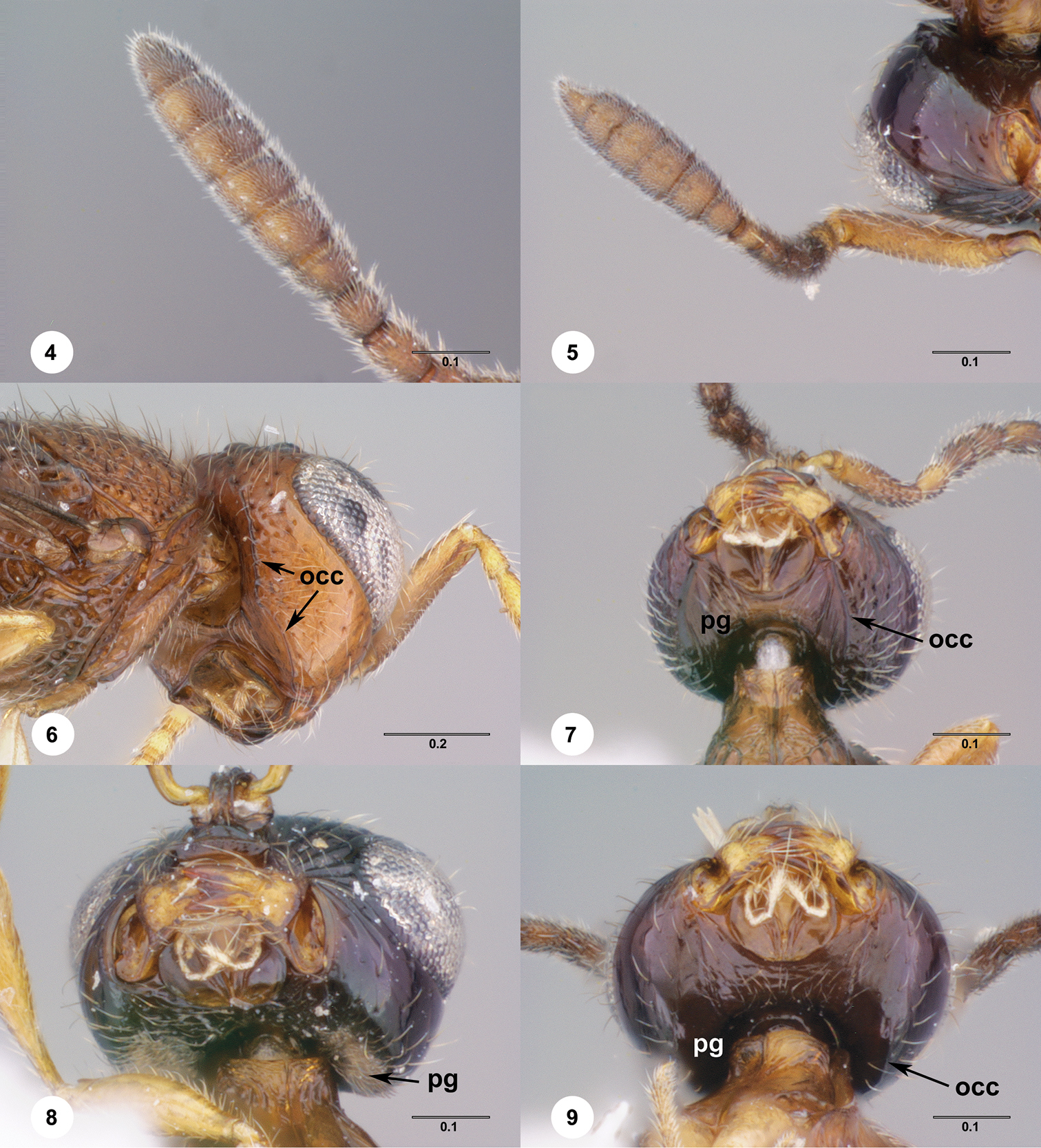
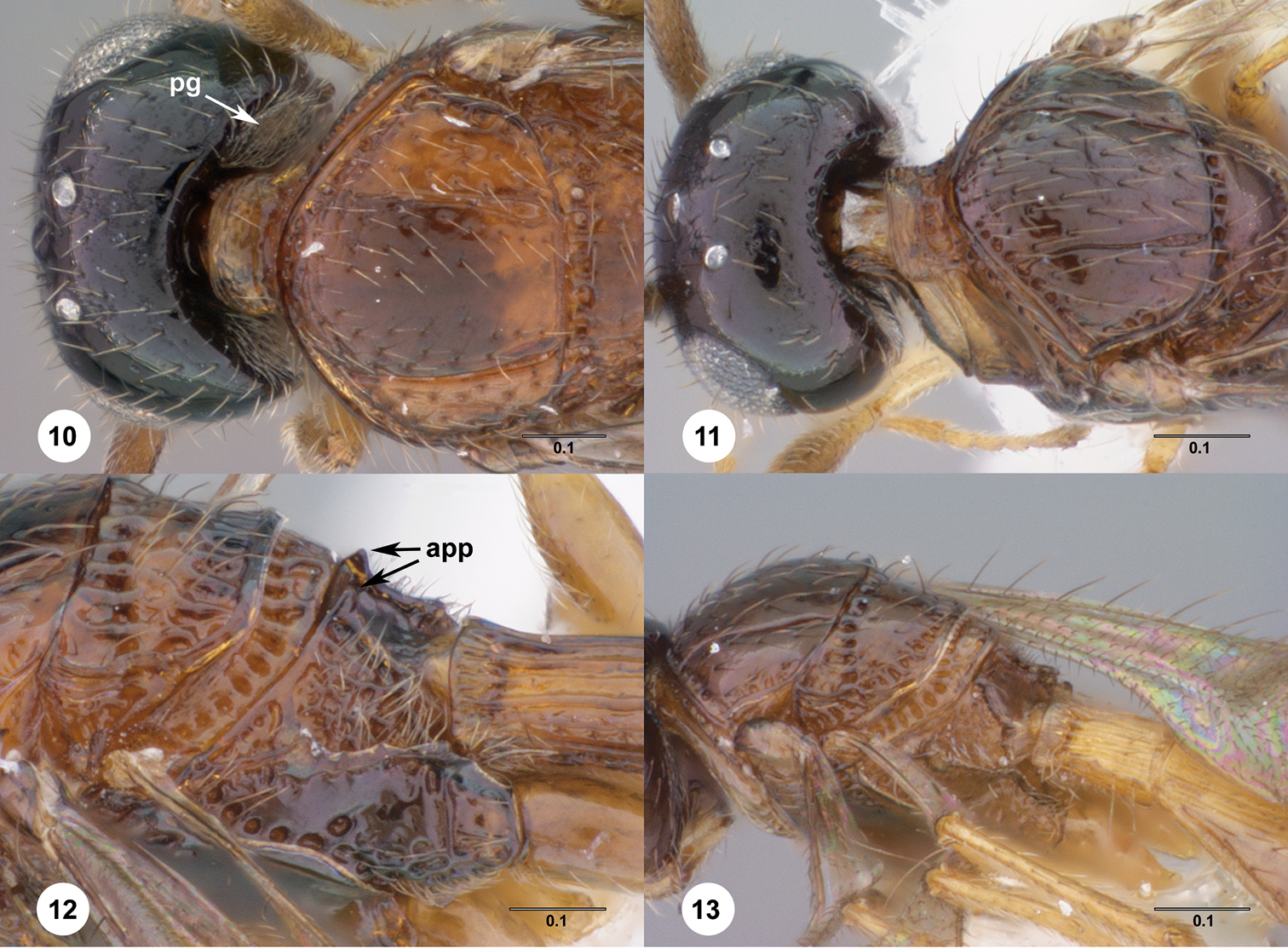
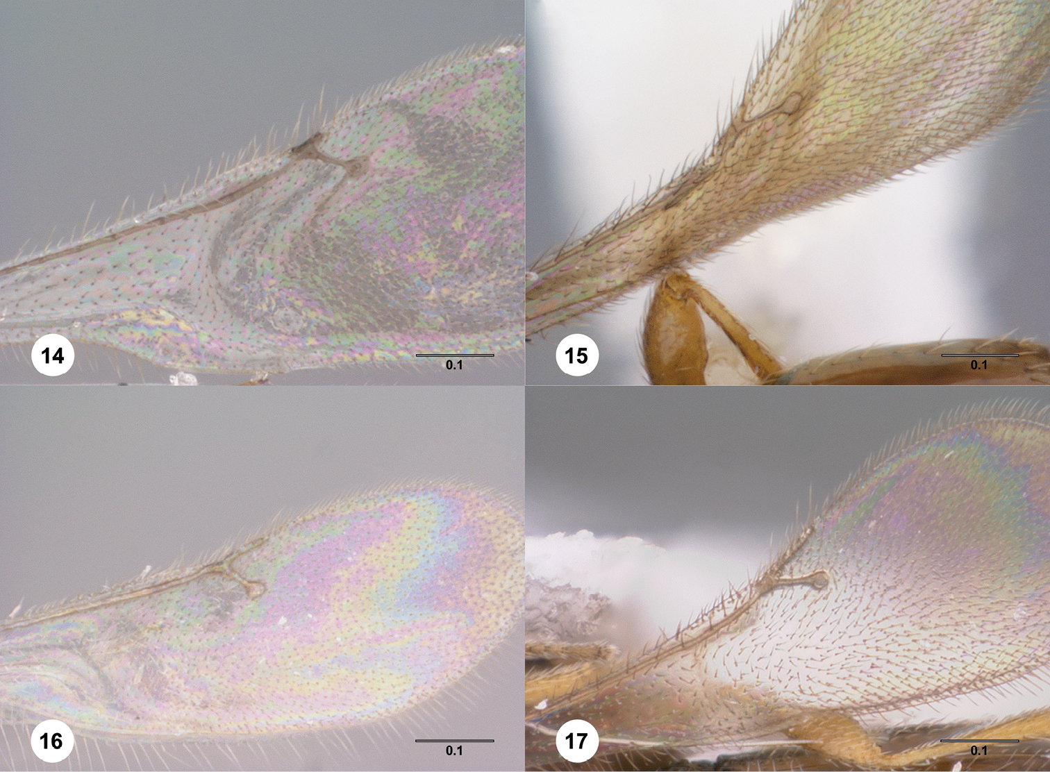
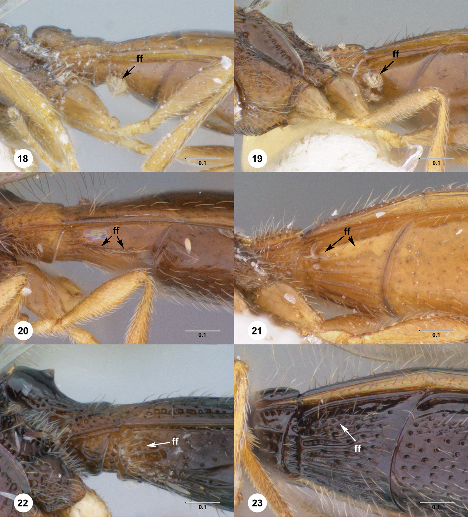
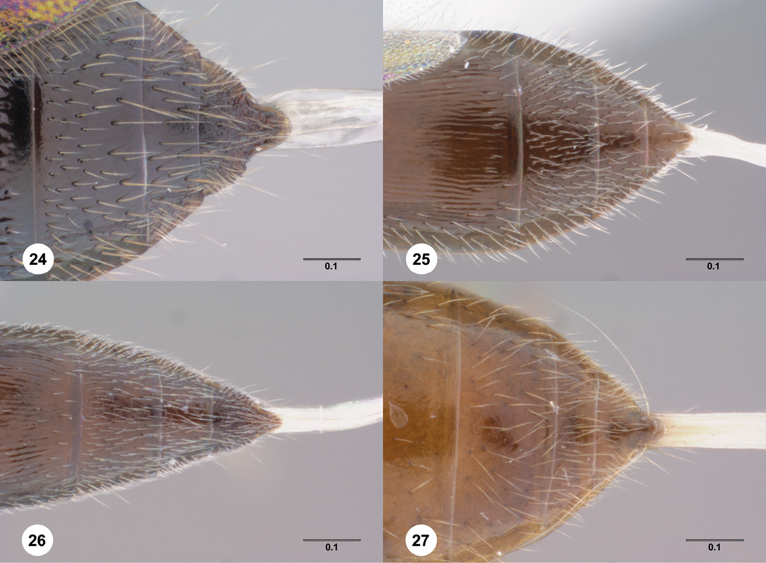
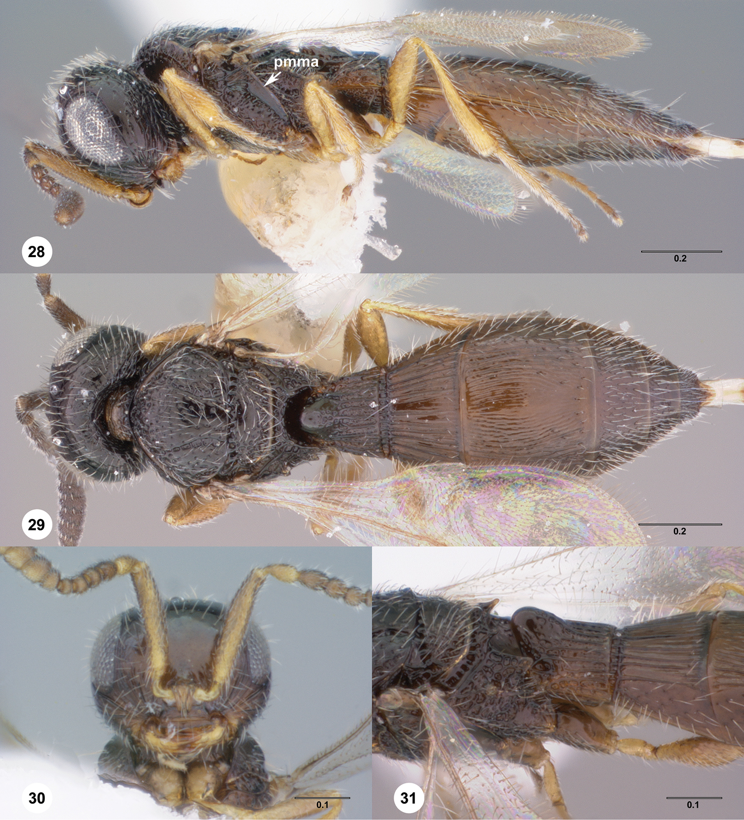
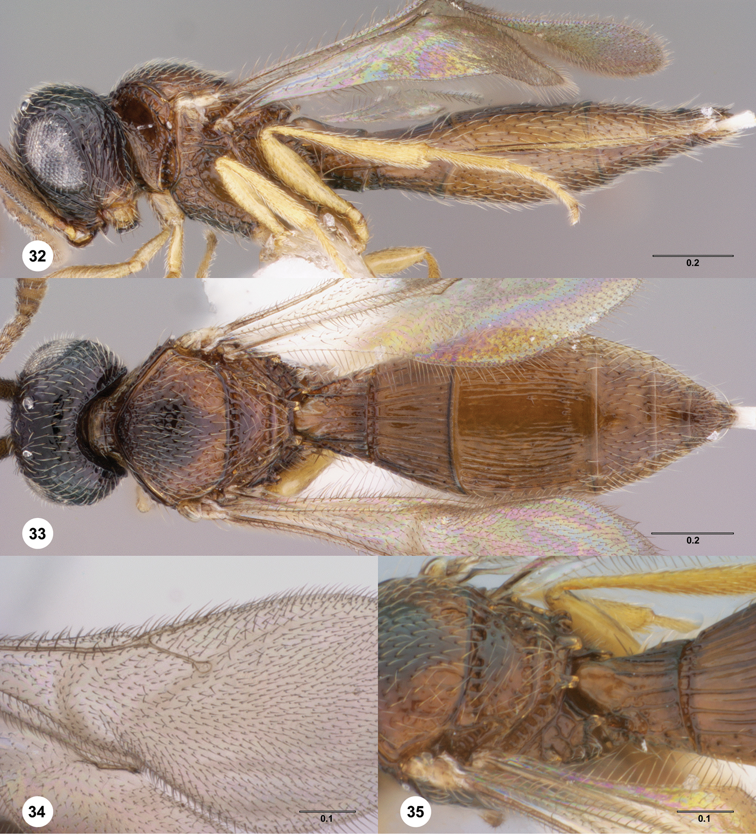
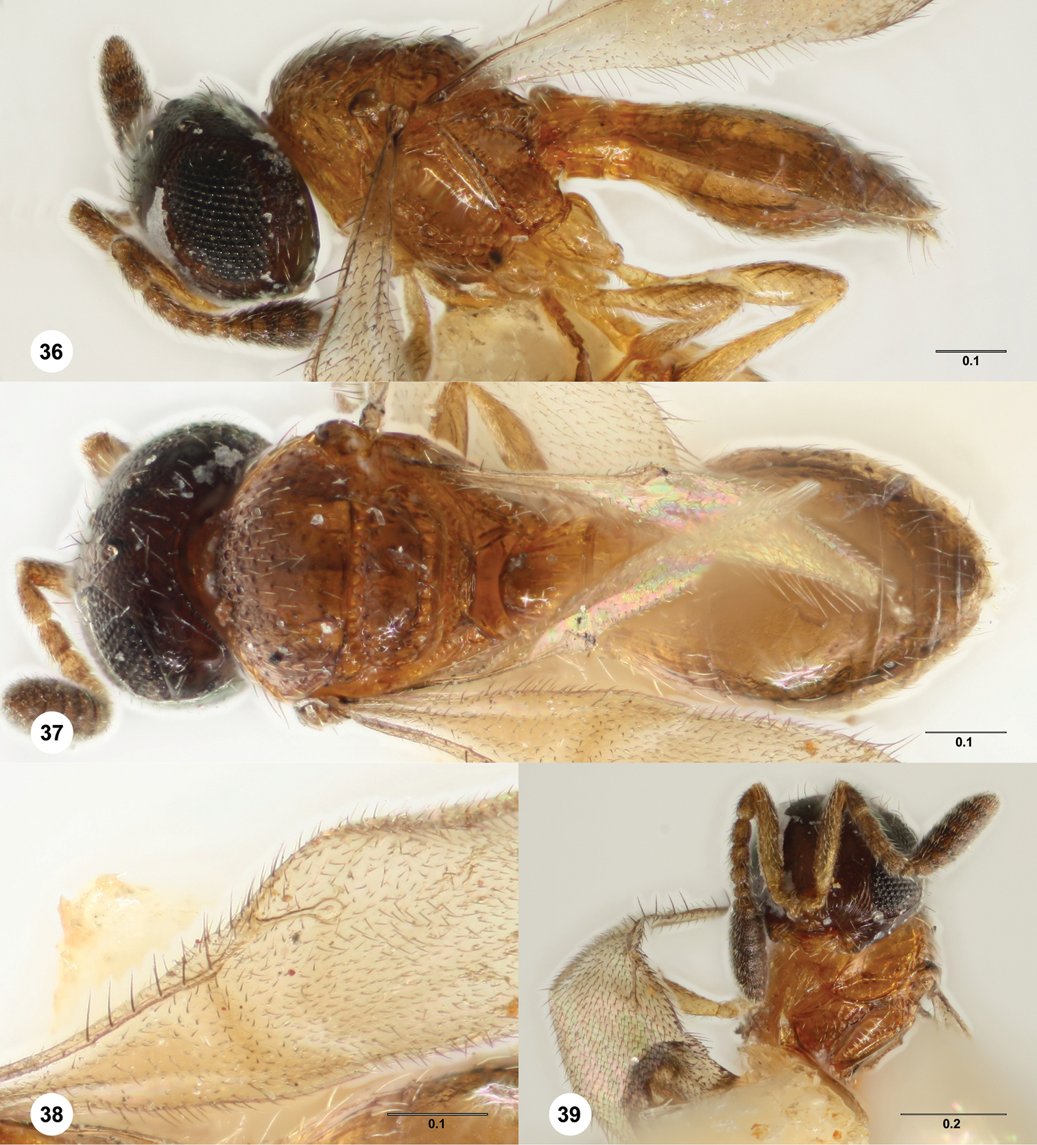
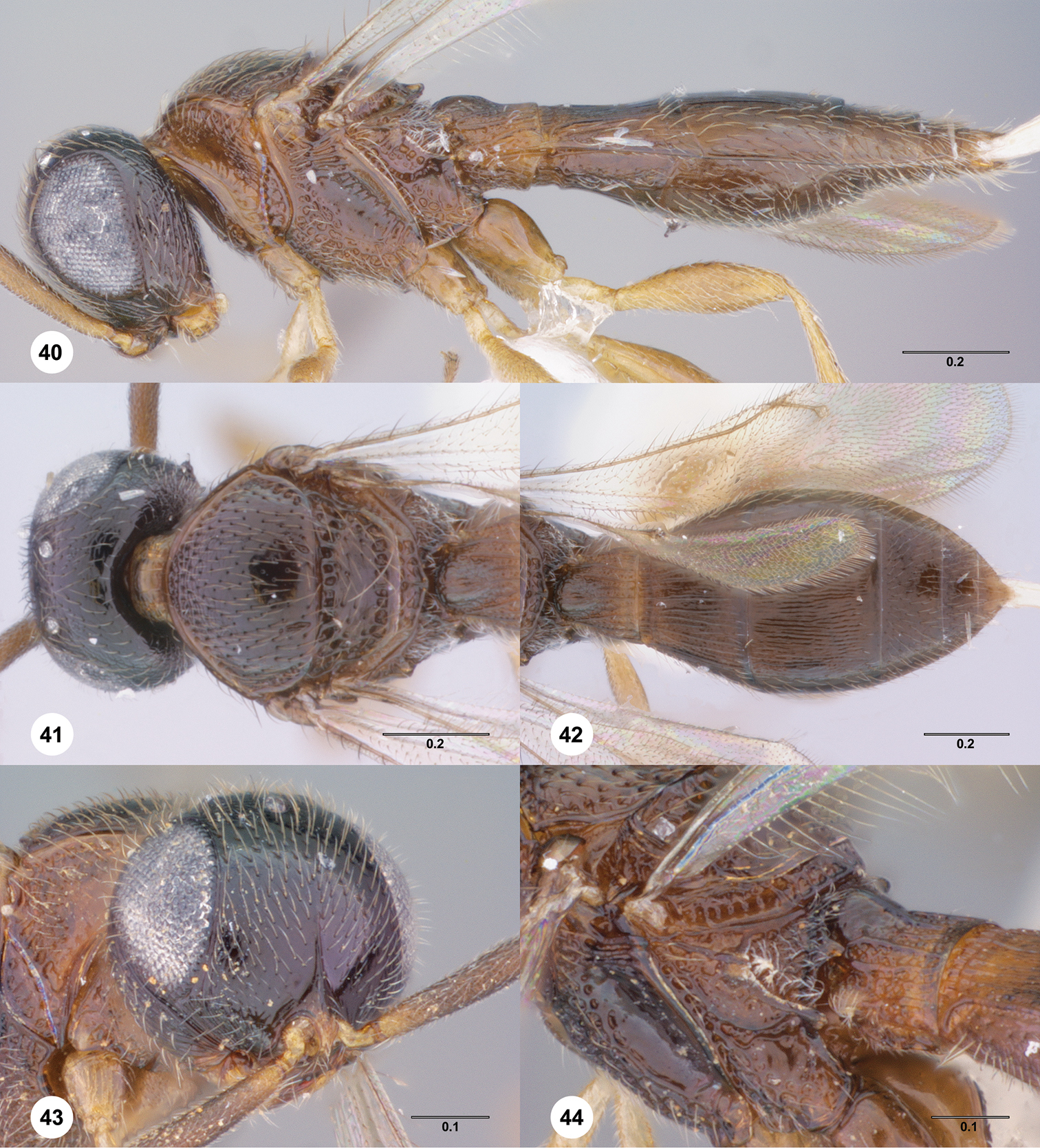
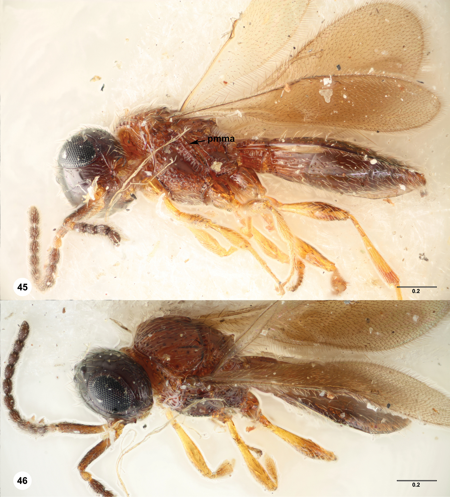
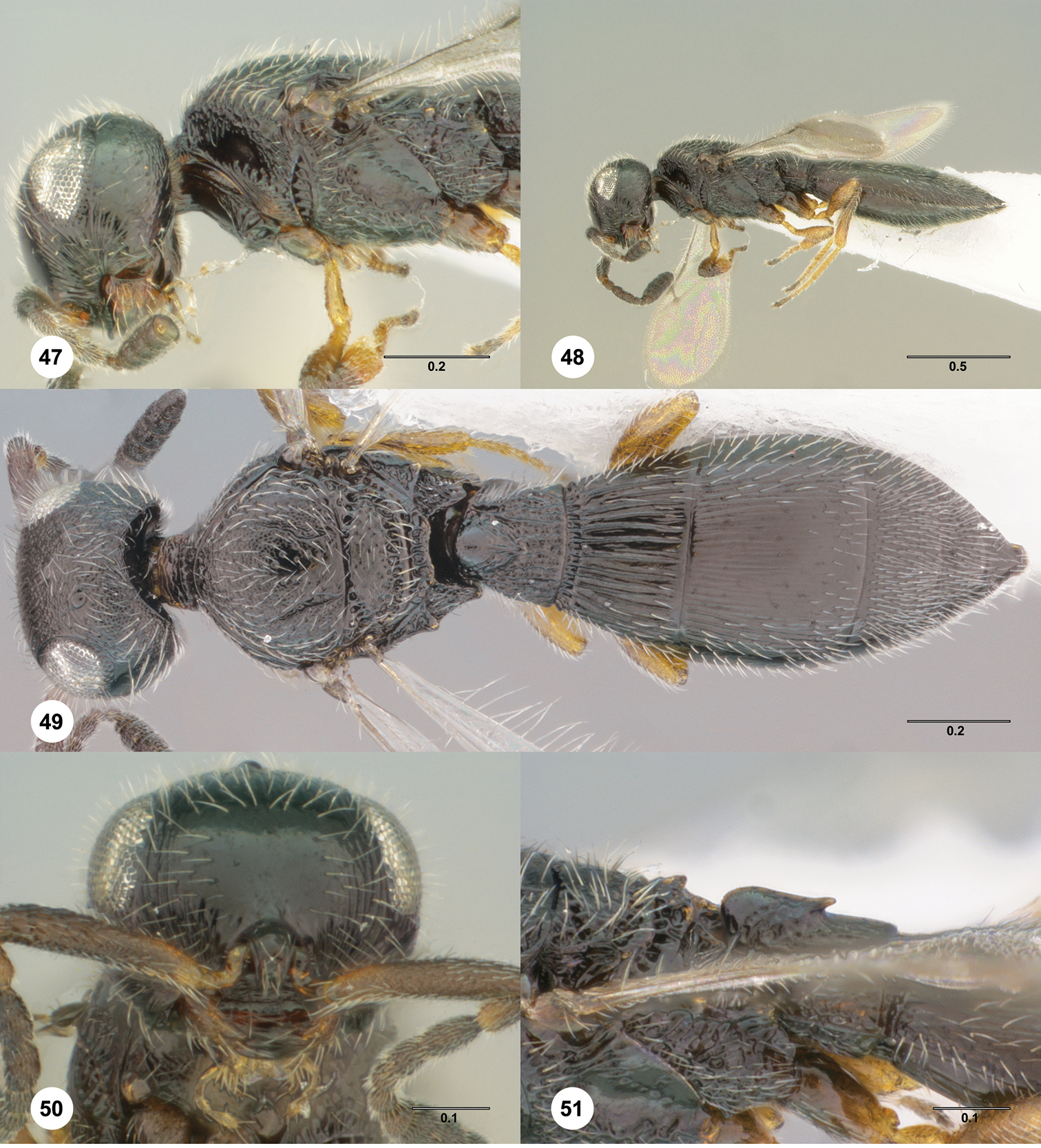
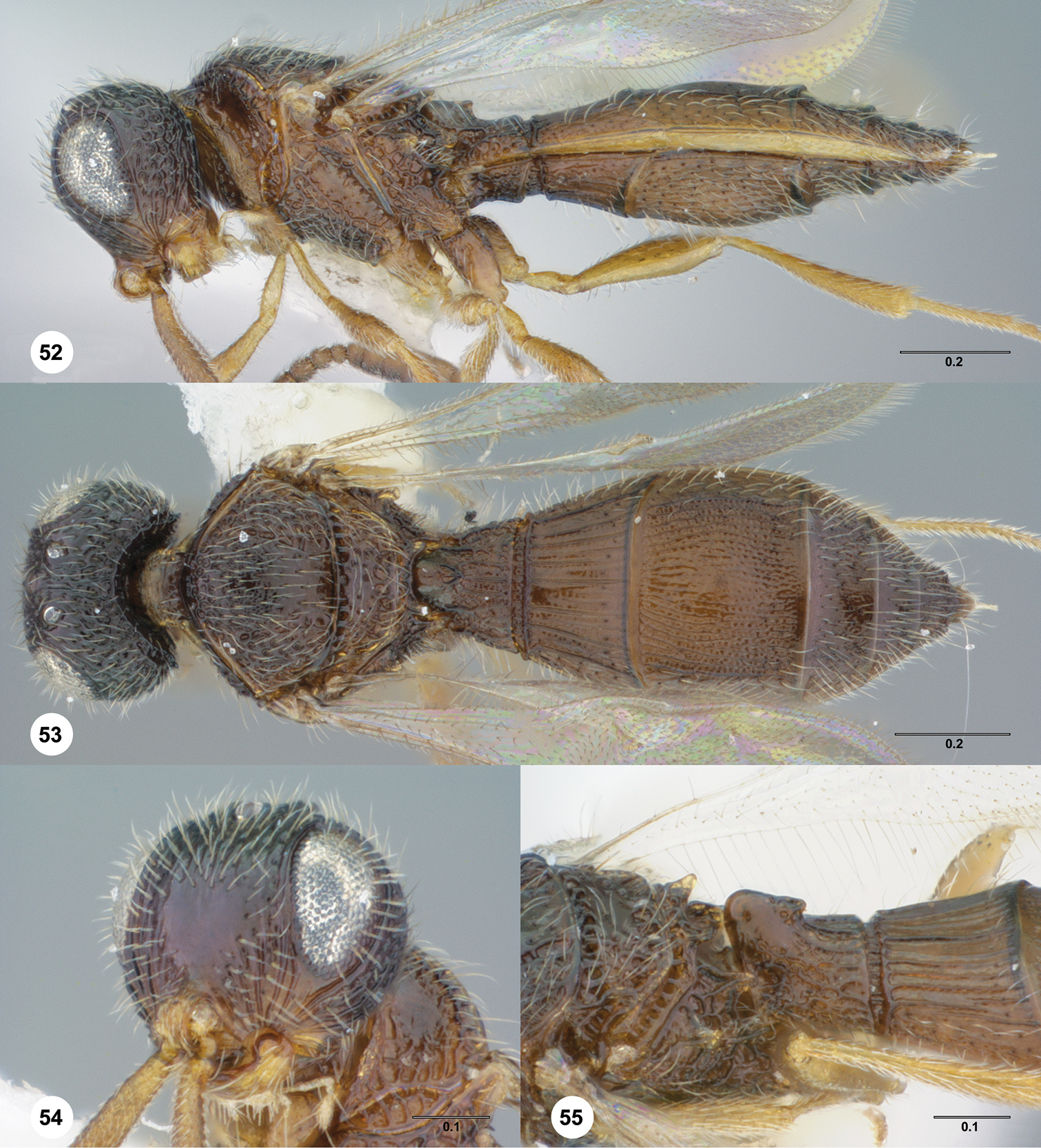
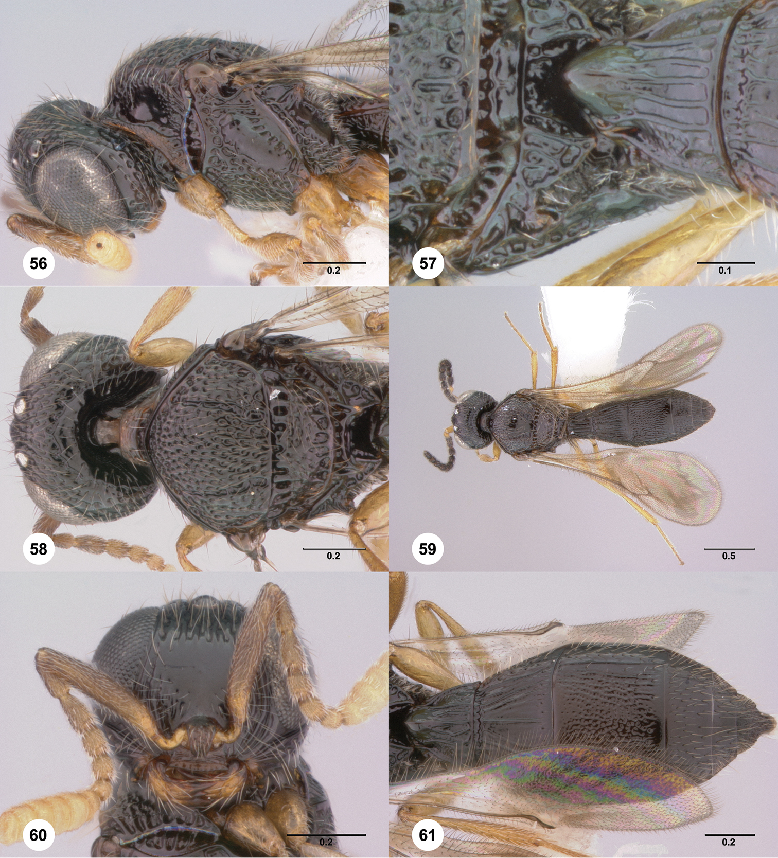
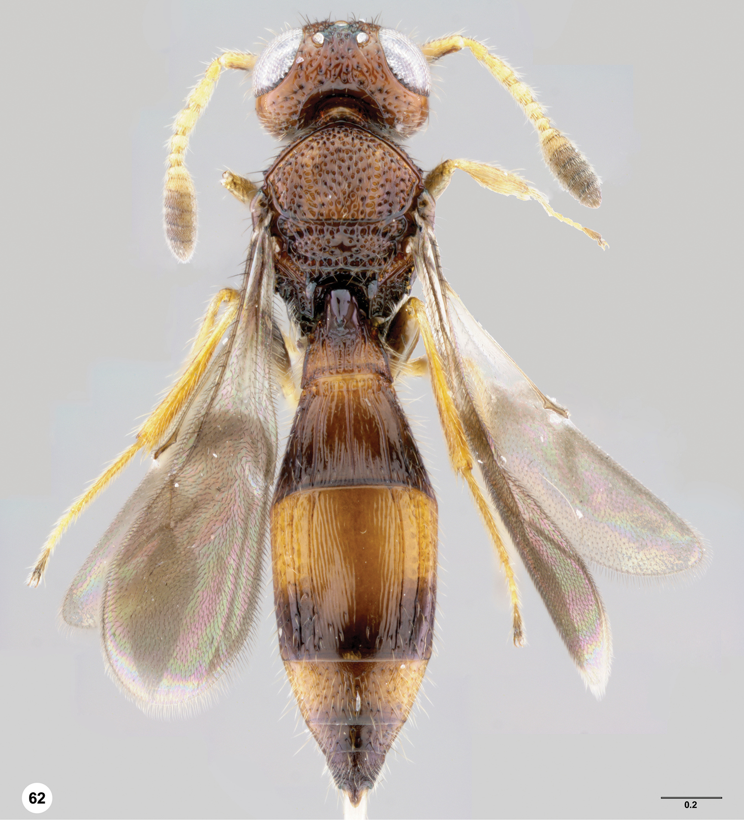
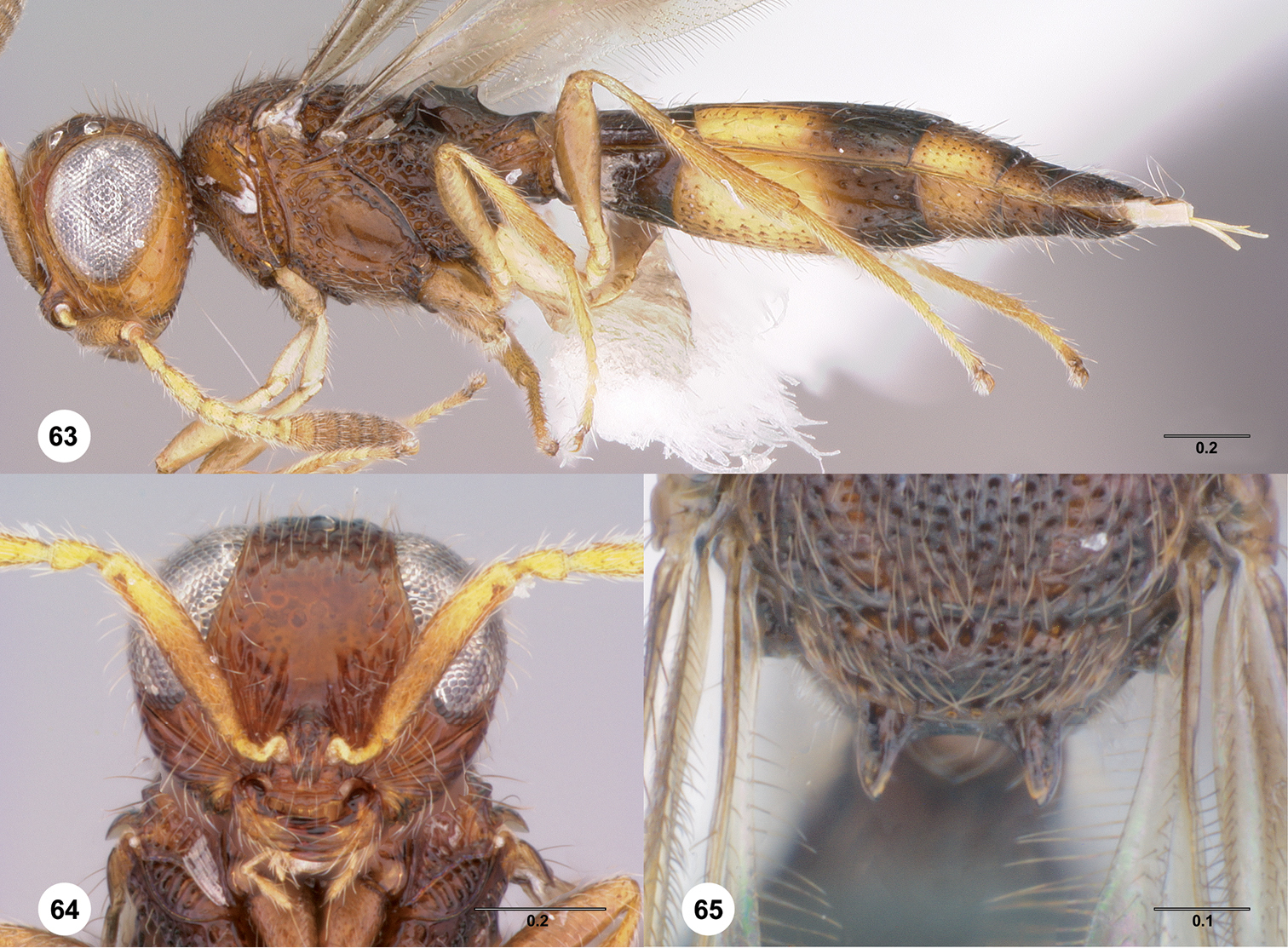
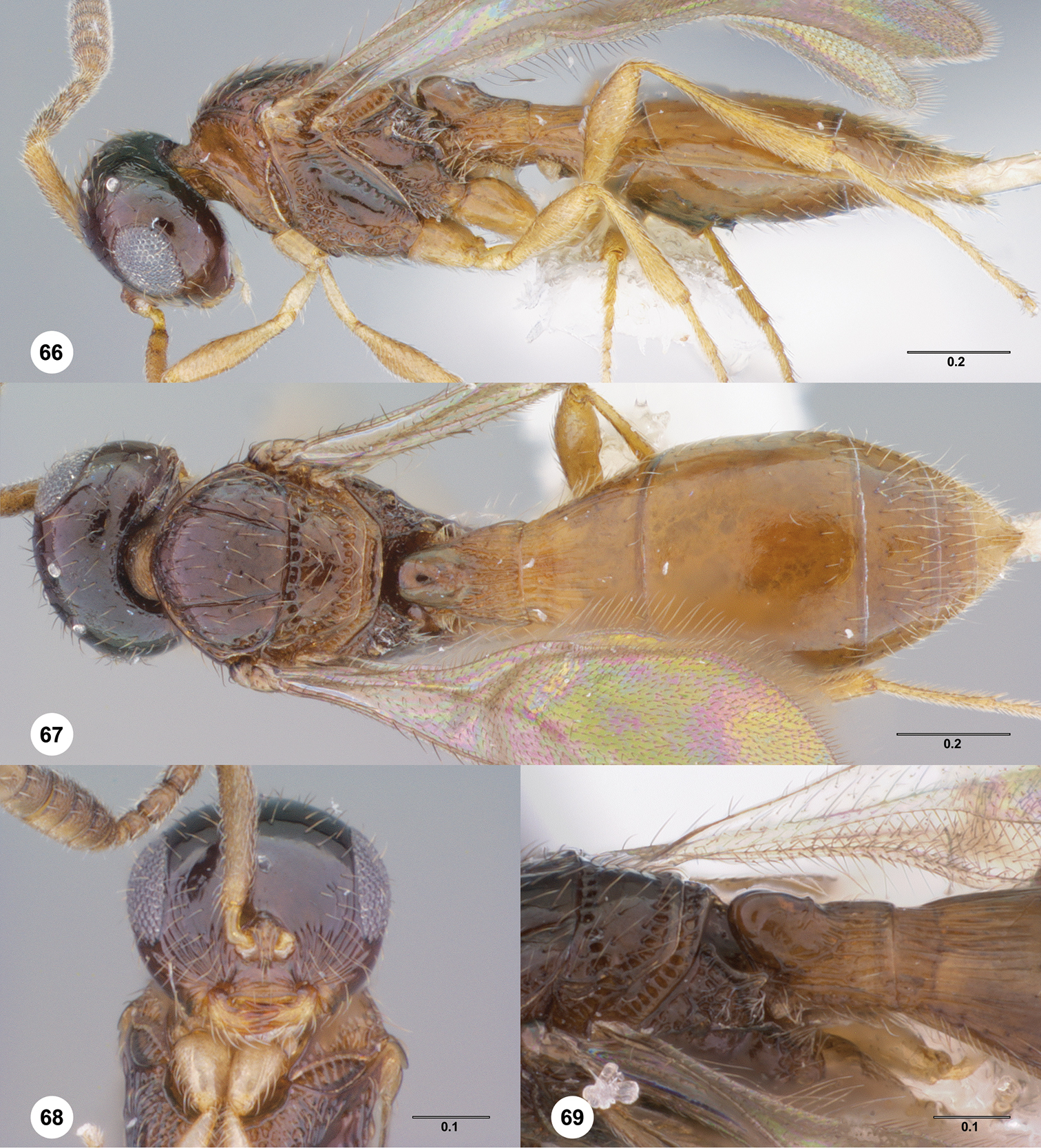
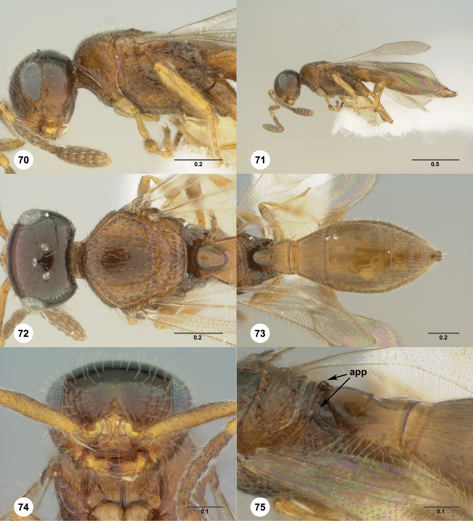
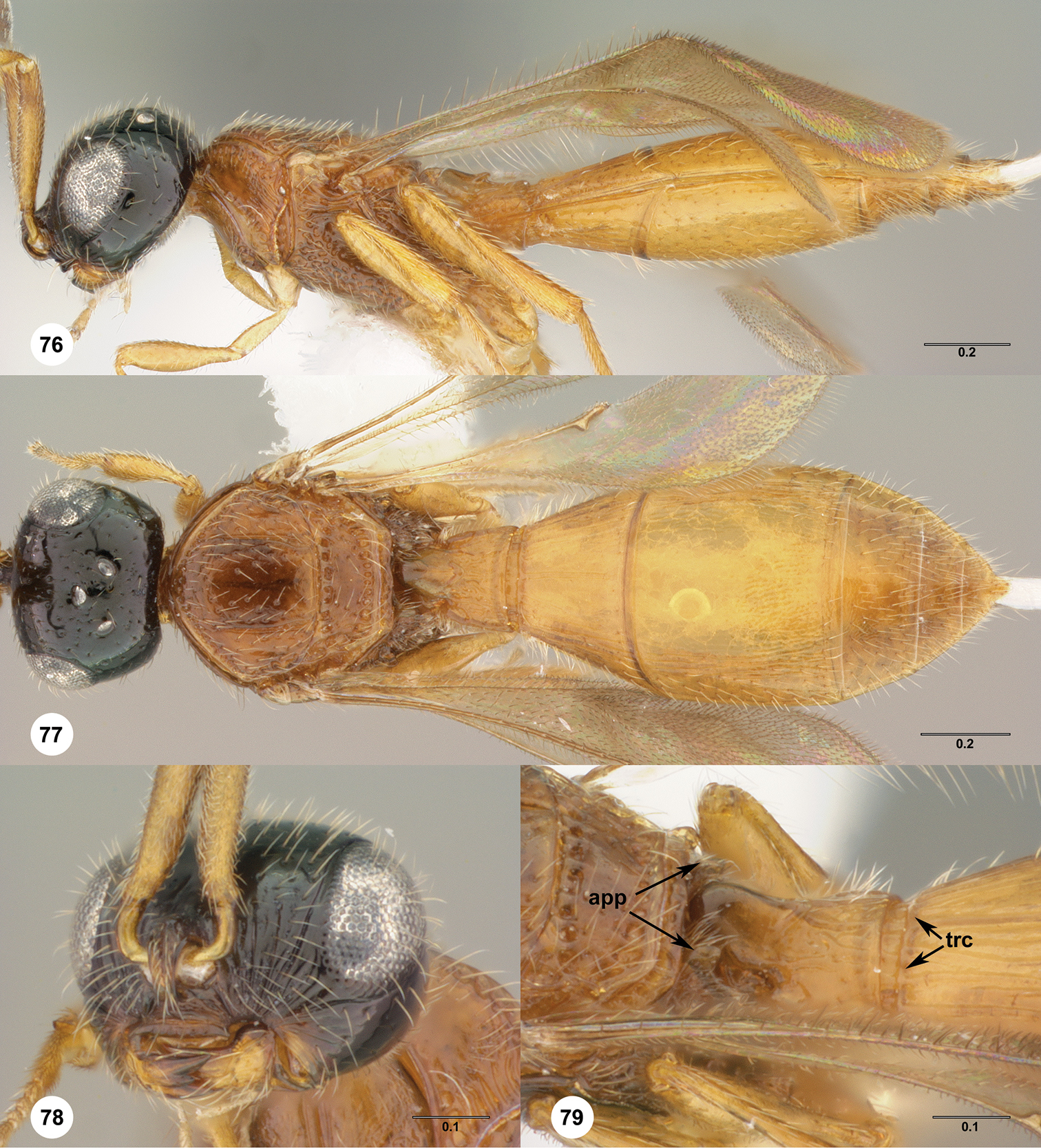
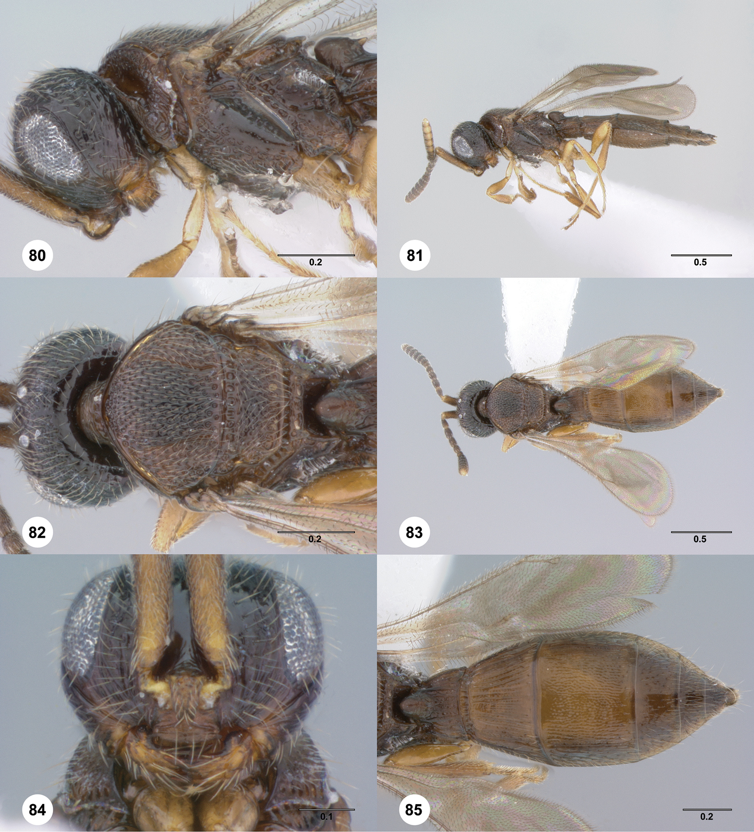
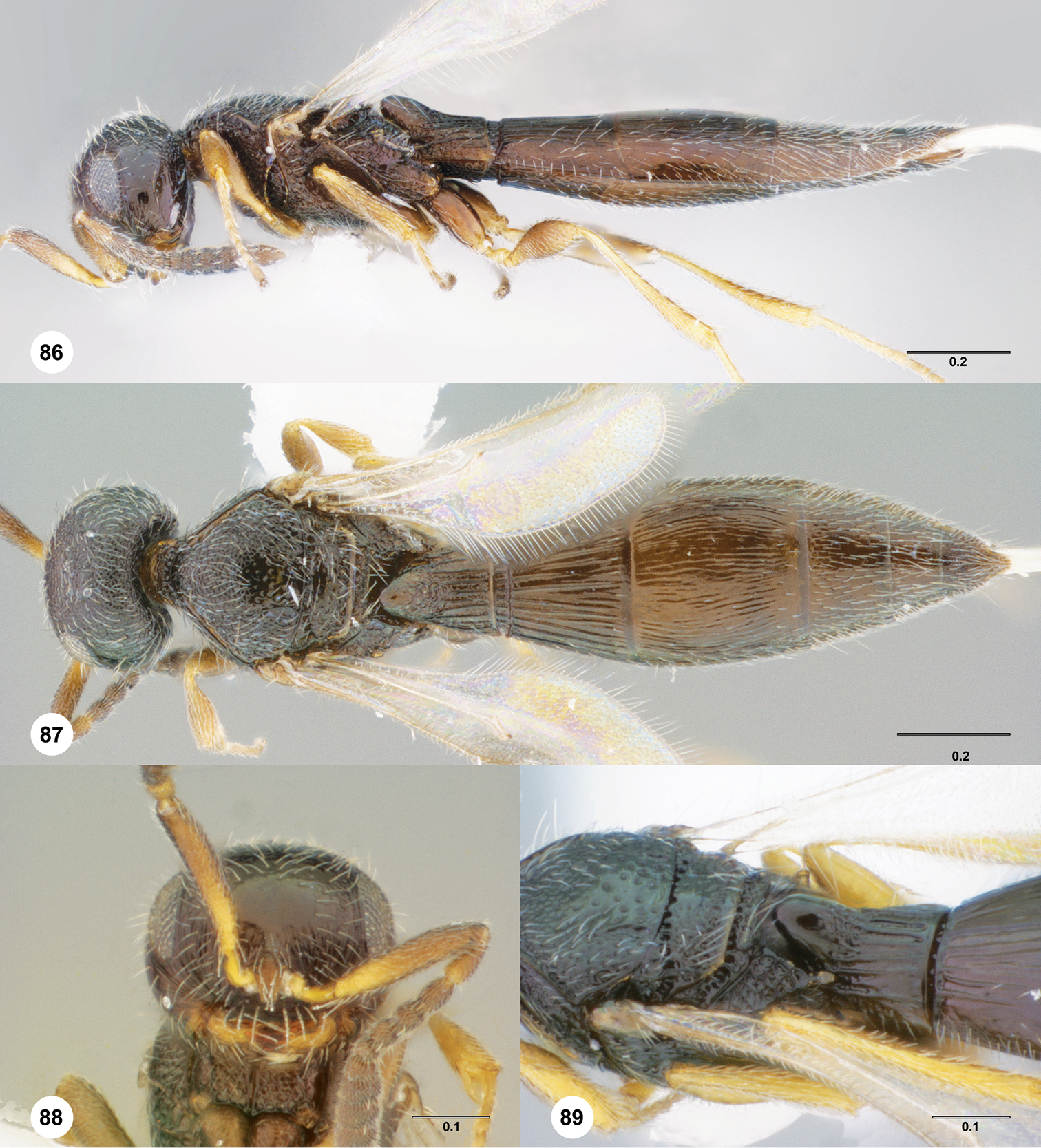
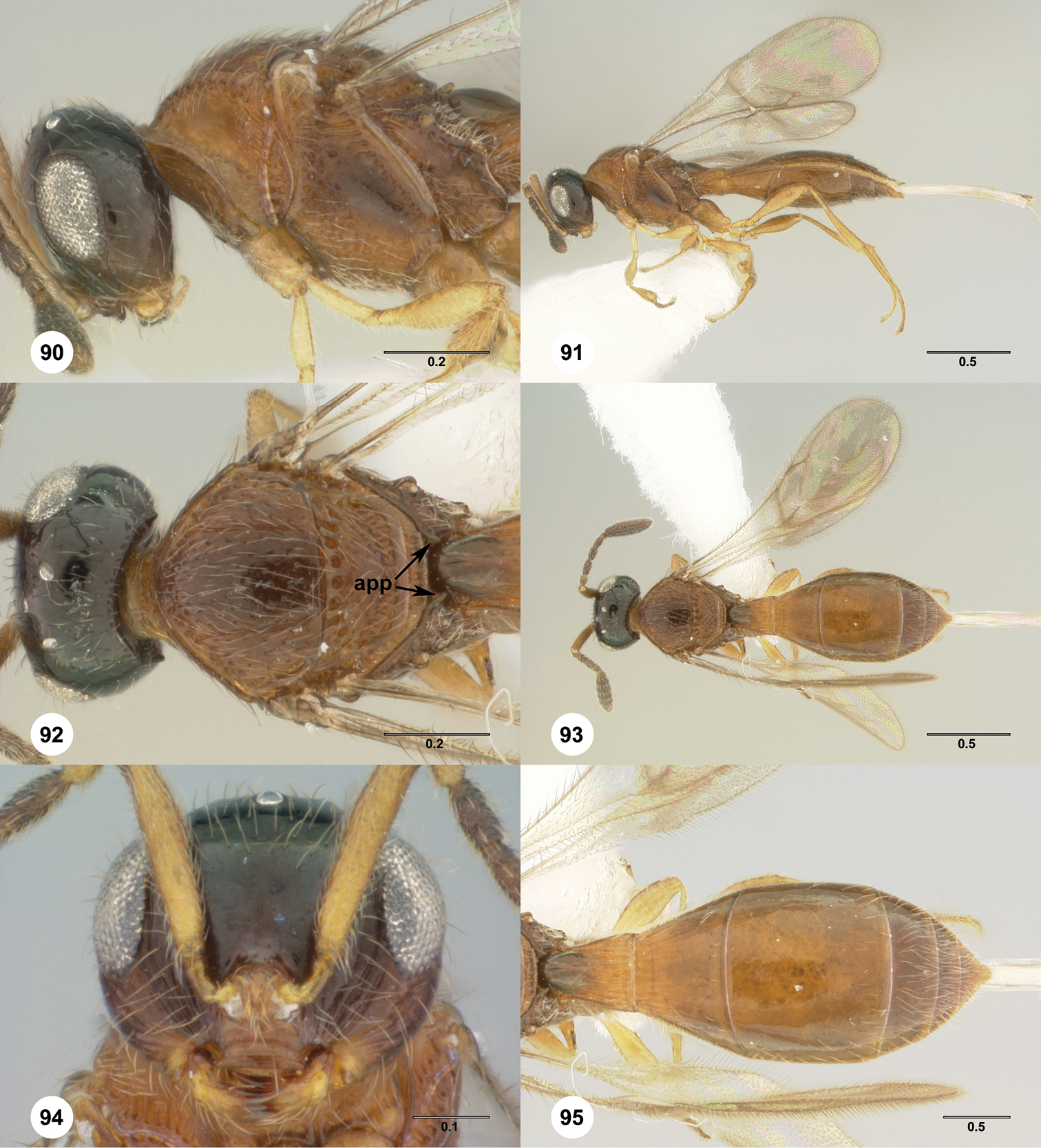
Thanks to K. van Achterberg, M. Buffington, R. Danielsson, T. Nuhn, S. van Noort, and M. Sharkey (Thai specimens collected under NSF grant No. DEB-0542864) for the loans of material for this study, to A. Polaszek and V. Blagoderov for help with imaging type material at BMNH, and to L. Musetti, J. Cora, and S. Hemly for critical assistance with specimen handling, software and databasing. This material is based upon work supported in part by the National Science Foundation under grant No. DEB-0614764 to N.F. Johnson and A.D.Austin.
1 http://biocol.org/urn:lsid:biocol.org:col:1008
2 http://biocol.org/urn:lsid:biocol.org:col:1009
3 http://biocol.org/urn:lsid:biocol.org:col:1010
4 http://biocol.org/urn:lsid:biocol.org:col:1011
5 http://biocol.org/urn:lsid:biocol.org:col:1012
6 http://biocol.org/urn:lsid:biocol.org:col:33357
7 http://biocol.org/urn:lsid:biocol.org:col:33791
8 http://biocol.org/urn:lsid:biocol.org:col:33943
9 http://biocol.org/urn:lsid:biocol.org:col:33864
10 http://biocol.org/urn:lsid:biocol.org:col:1014
11 http://biocol.org/urn:lsid:biocol.org:col:1016
12 http://biocol.org/urn:lsid:biocol.org:col:34212
13 http://biocol.org/urn:lsid:biocol.org:col:1018
14 http://biocol.org/urn:lsid:biocol.org:col:1019
15 http://morphbank.net/?id=796511
16 http://hol.osu.edu/map-large.html?id=303979
17 http://morphbank.net/?id=796513
18 http://hol.osu.edu/map-large.html?id=5063
19 http://morphbank.net/?id=796512
20 http://hol.osu.edu/map-large.html?id=5066
21 http://morphbank.net/?id=796514
22 http://morphbank.net/?id=796515
23 http://hol.osu.edu/map-large.html?id=5076
24 http://morphbank.net/?id=796516
25 http://hol.osu.edu/map-large.html?id=5081
26 http://hol.osu.edu/map-full.html?id=813379
27 http://hol.osu.edu/map-full.html?id=315505
28 http://morphbank.net/?id=796716
29 http://hol.osu.edu/map-large.html?id=4931
30 http://morphbank.net/?id=796717
31 http://hol.osu.edu/map-large.html?id=305675
32 http://morphbank.net/?id=796718
33 http://hol.osu.edu/map-large.html?id=303879
34 http://morphbank.net/?id=796719
35 http://hol.osu.edu/map-large.html?id=303882
36 http://morphbank.net/?id=796720
37 http://hol.osu.edu/map-large.html?id=303880
38 http://morphbank.net/?id=796721
39 http://hol.osu.edu/map-large.html?id=303975
40 http://morphbank.net/?id=796722
41 http://hol.osu.edu/map-large.html?id=303974
42 http://morphbank.net/?id=796723
43 http://hol.osu.edu/map-large.html?id=303881
44 http://lsid.tdwg.org/urn:lsid:biosci.ohio-state.edu:osuc_pubs:131
45 http://lsid.tdwg.org/urn:lsid:biosci.ohio-state.edu:osuc_pubs:21126
46 http://lsid.tdwg.org/urn:lsid:biosci.ohio-state.edu:osuc_pubs:460
47 doi: 10.1093/nar/gkh340
48 http://lsid.tdwg.org/urn:lsid:biosci.ohio-state.edu:osuc_pubs:353
49 http://lsid.tdwg.org/urn:lsid:biosci.ohio-state.edu:osuc_pubs:339
50 doi: 10.1007/s00239-004-0246-x
51 doi: 10.1093/molbev/msi152
52 doi: 10.1111/j.1096-0031.2008.00217.x
53 http://lsid.tdwg.org/urn:lsid:biosci.ohio-state.edu:osuc_pubs:22787
54 http://lsid.tdwg.org/urn:lsid:biosci.ohio-state.edu:osuc_pubs:21725
55 http://lsid.tdwg.org/urn:lsid:biosci.ohio-state.edu:osuc_pubs:23446
56 http://lsid.tdwg.org/urn:lsid:biosci.ohio-state.edu:osuc_pubs:386
57 http://lsid.tdwg.org/urn:lsid:biosci.ohio-state.edu:osuc_pubs:387
58 http://lsid.tdwg.org/urn:lsid:biosci.ohio-state.edu:osuc_pubs:310
59 http://lsid.tdwg.org/urn:lsid:biosci.ohio-state.edu:osuc_pubs:22163
60 http://lsid.tdwg.org/urn:lsid:biosci.ohio-state.edu:osuc_pubs:349
61 http://lsid.tdwg.org/urn:lsid:biosci.ohio-state.edu:osuc_pubs:342
62 http://lsid.tdwg.org/urn:lsid:biosci.ohio-state.edu:osuc_pubs:311
63 http://lsid.tdwg.org/urn:lsid:biosci.ohio-state.edu:osuc_pubs:12273
64 http://lsid.tdwg.org/urn:lsid:biosci.ohio-state.edu:osuc_pubs:312
65 http://lsid.tdwg.org/urn:lsid:biosci.ohio-state.edu:osuc_pubs:7039
66 http://www.mapress.com/zootaxa/2007f/zt01571p078.pdf
67 http://lsid.tdwg.org/urn:lsid:biosci.ohio-state.edu:osuc_pubs:21247
68 http://lsid.tdwg.org/urn:lsid:biosci.ohio-state.edu:osuc_pubs:336
69 doi: 10.1016/j.ympev.2006.02.007
70 http://lsid.tdwg.org/urn:lsid:biosci.ohio-state.edu:osuc_pubs:20959
71 http://lsid.tdwg.org/urn:lsid:biosci.ohio-state.edu:osuc_pubs:21327
72 http://lsid.tdwg.org/urn:lsid:biosci.ohio-state.edu:osuc_pubs:379
73 http://lsid.tdwg.org/urn:lsid:biosci.ohio-state.edu:osuc_pubs:22369
74 http://lsid.tdwg.org/urn:lsid:biosci.ohio-state.edu:osuc_pubs:23390
75 http://lsid.tdwg.org/urn:lsid:biosci.ohio-state.edu:osuc_pubs:23583
76 http://lsid.tdwg.org/urn:lsid:biosci.ohio-state.edu:osuc_pubs:23802
77 doi: 10.1371/journal.pone.0015991
78 http://lsid.tdwg.org/urn:lsid:biosci.ohio-state.edu:osuc_pubs:23164
79 http://morphbank.net/?id=796518
80 http://morphbank.net/?id=796519
81 http://morphbank.net/?id=796520
82 http://morphbank.net/?id=796521
83 http://morphbank.net/?id=796522
84 http://morphbank.net/?id=796523
85 http://morphbank.net/?id=796524
86 http://morphbank.net/?id=796512
87 http://morphbank.net/?id=796525
88 http://morphbank.net/?id=796515
89 http://morphbank.net/?id=796526
90 http://morphbank.net/?id=813380
91 http://morphbank.net/?id=796724
92 http://morphbank.net/?id=796725
93 http://morphbank.net/?id=796726
94 http://morphbank.net/?id=796727
95 http://morphbank.net/?id=796728
96 http://morphbank.net/?id=796729
97 http://morphbank.net/?id=796730
URI table of HAO morphological terms. (doi: 10.3897/JHR.34.4714.app) File format: Microsoft Word Open XML Document (docx).
List of morphological characters and character states mapped onto the strict consensus phylogeny of combined morphological and molecular data. (doi: 10.3897/JHR.34.4714.app2) File format: Microsoft Word Open XML Document (docx).
Strict consensus phylogeny of combined morphological and molecular data with Partitioned Bremer Support assigned to resolved nodes. Partition 1: morphology; Partition 2: 28S; Partition 3: 18S; Partition 4: CO1. (doi: 10.3897/JHR.34.4714.app3) File format: Portable Network Graphics (png).
Tables of CUIDs for DNA voucher specimens and Genbank accession numbers for sequences used in phylogenetic analyses. (doi: 10.3897/JHR.34.4714.app4) File format: Microsoft Word Open XML Document (docx).
Interpolated matrix used for phylogenetic analysis. Partition 1: morphology; Partition 2: 28S; Partition 3: 18S; Partition 4: CO1. (doi: 10.3897/JHR.34.4714.app5) File format: TNT Matrix File (TNT).