Monograph |
|
Corresponding author: Ovidiu Alin Popovici ( popovici_alin_ovidiu@yahoo.com ) Academic editor: Miles Zhang
© 2022 Ovidiu Alin Popovici, Lubomir Masner, Zachary Lahey, Elijah Talamas.
This is an open access article distributed under the terms of the CC0 Public Domain Dedication.
Citation:
Popovici OA, Masner L, Lahey Z, Talamas E (2022) Revision of the Palearctic species of Fidiobia Ashmead (Hymenoptera, Platygastroidea). Journal of Hymenoptera Research 92: 23-144. https://doi.org/10.3897/jhr.92.85040
|
Abstract
The Palearctic species of the genus Fidiobia are revised, seventeen new species are described (F. bohemica sp. nov.; F. brevialis sp. nov.; F. communis sp. nov.; F. gallica sp. nov.; F. hirta sp. nov.; F. insoonae sp. nov.; F. lisenchiae sp. nov.; F. longiclava sp. nov.; F. nipponica sp. nov.; F. platystasioides sp. nov.; F. politoides sp. nov.; F. pronotatoides sp. nov.; F. roatai sp. nov.; F. rugosifronsoides sp. nov.; F. sashai sp. nov.; F. tripotini sp. nov.; F. vladlubomiri sp. nov.), and eleven species (F. brevinotaula Veenakumari et al., 2018; F. filicornis Buhl, 2014; F. flaviabdominalis Veenakumari et al., 2018; F. hispanica Popovici & Buhl, 2010; F. hofferi Kozlov, 1978; F. polita Buhl, 1998; F. pronotata Szabó, 1958; F. rugosifrons Crawford, 1916; F. striatitergitis (Szabó, 1962); F. synergorum (Kieffer, 1921); F. vanharteni Buhl, 2010) are redescribed. A key for identification of females and distributional data for each species are provided. Brachypterous specimens are reported for F. rugosifrons and F. hofferi. Fidiobia gordoni Popovici & Buhl, 2010 is treated as a junior synonym of Fidiobia striatitergitis (Szabó, 1962).
Keywords
α-taxonomy, egg-parasitoids, new species, Platygastridae, Sceliotrachelinae
Introduction
Fidiobia Ashmead, 1894 is one of the “classical” genera of Platygastridae, originally described as monotypic, with F. flavipes Ashmead, 1894 as the type species.
Taxonomic history
The next important step in the taxonomy of Fidiobia was made by
Until 1989, only ten species of Fidiobia had been formally described (Table
Species of Fidiobia described prior to this study and their distribution.
| Nr. crt. | Species | Author(s) | Year | Biogeographical regions |
|---|---|---|---|---|
| 1 | F. benjamini | (Nixon) | 1969 | Afrotropical |
| 2 | F. danielssoni | Buhl | 2001 | |
| 3 | F. tanzaniana | Buhl | 2010 | |
| 4 | F. vanharteni | Buhl | 2010 | |
| 5 | F. zebra | Buhl | 2010 | |
| 6 | F. filicornis | Buhl | 2014 | |
| 7 | F. semirufa | Buhl | 2014 | |
| 8 | F. tschirnhausi | Buhl | 2014 | |
| 9 | F. celeritas | van Noort and Lahey | 2021 | |
| 10 | F. synergorum | (Kieffer) | 1921 | Palearctic |
| 11 | F. pronotata | Szabó | 1958 | |
| 12 | F. hofferi | Kozlov | 1978 | |
| 13 | F. polita | Buhl | 1998 | |
| 14 | F. gordoni | Popovici and Buhl | 2010 | |
| 15 | F. hispanica | Popovici and Buhl | 2010 | |
| 16 | F. flavipes | Ashmead | 1894 | Nearctic |
| 17 | F. drakei | (Oglobin) | 1944 | |
| 18 | F. rugosifrons | Crawford | 1916 | Holarctic |
| 19 | F. bonariensis | (Brèthes) | 1916 | Neotropical |
| 20 | F. citri | (Nixon) | 1969 | |
| 21 | F. asina | (Loiácono) | 1982 | |
| 22 | F. dominica | Evans and Peña | 2005 | |
| 23 | F. flava | Buhl | 2011 | |
| 24 | F. semistriata | Buhl et al. | 2009 | Oriental |
| 25 | F. nagarajae | Veenakumari et al. | 2012 | |
| 26 | F. virakthamati | Veenakumari et al. | 2012 | |
| 27 | F. brevinotaula | Veenakumari et al. | 2018 | |
| 28 | F. carinata | Veenakumari et al. | 2018 | |
| 29 | F. crocea | Veenakumari et al. | 2018 | |
| 30 | F. dantela | Veenakumari et al. | 2018 | |
| 31 | F. decora | Veenakumari et al. | 2018 | |
| 32 | F. doddi | Veenakumari et al. | 2018 | |
| 33 | F. flaviabdominalis | Veenakumari et al. | 2018 | |
| 34 | F. flavifrons | Veenakumari et al. | 2018 | |
| 35 | F. fusca | Veenakumari et al. | 2018 | |
| 36 | F. galben | Veenakumari et al. | 2018 | |
| 37 | F. hima | Veenakumari et al. | 2018 | |
| 38 | F. leptidantela | Veenakumari et al. | 2018 | |
| 39 | F. longiabdominalis | Veenakumari et al. | 2018 | |
| 40 | F. multicarinata | Veenakumari et al. | 2018 | |
| 41 | F. nandi | Veenakumari et al. | 2018 | |
| 42 | F. nilgiriensis | Veenakumari et al. | 2018 | |
| 43 | F. prashanthi | Veenakumari et al. | 2018 | |
| 44 | F. punyakoti | Veenakumari et al. | 2018 | |
| 45 | F. setosa | Veenakumari et al. | 2018 | |
| 46 | F. striatipleura | Veenakumari et al. | 2018 | |
| 47 | F. szaboi | Veenakumari et al. | 2018 | |
| 48 | F. vandu | Veenakumari et al. | 2018 |
This paper is only a small part of a large and ambitious project concerning the revision of the world fauna of Fidiobia, a genus of beetle egg parasitoids.
Materials and methods
Specimens
Taxonomic studies are greatly informed by the examination of large numbers of specimens to determine morphological variation and geographic distribution, and to associate conspecific males and females. Fidiobia is not a rare genus, but the number of specimens in European collections is typically very small. We believe this is partly because knowledge about the biology of the genus is lacking, which hinders the development of more efficient collecting strategies. For example, some genera that are now commonly collected (e.g., Baeoneurella Dodd, Tiphodytes Bradley, Baryconus Förster) were once considered rare. Now that the biology of some of their species has been elucidated, they can be easily captured with appropriate methods in certain habitats.
The specimens used in this study are deposited in the following institutions with the abbreviations (in bold) used in the text and the name of curators in parentheses:
ISNB Institut royal des Sciences naturelles de Belgique, Bruxelles (Yvonnick Gerard);
OPPC O.A. Popovici personal collection, stored in the Faculty of Biology, University ‘Al. I. Cuza’ Iasi, Romania;
Primary types
Images of the primary types of Fidiobia rugosifrons Crawford, 1916, were made available via the Museum of Biological Diversity database at The Ohio State University (https://mbd-db.osu.edu/hol/collecting_units/0eae5d1c-58ba-3aaa-e053-0100007f2cc9) by Prof. Dr. Norman Johnson. Primary types of Platyllotropa gallicola Szelényi, 1938 (
Collecting and preserving methods
The specimens used in this study were collected using a variety of methods. For each specimen, the collecting method, when known, is placed in parentheses using the following abbreviations:
LT light trap.
MT Malaise trap. For some species this method was very useful in obtaining a series of specimens.
SN sweep net. This method uses an entomological net with a circular or triangular frame. Specimens are extracted individually using an aspirator.
SS screen sweeping. This method uses an entomological net with a triangular frame (
TT conical trunk traps for collecting parasites of xylobionts (
YPT yellow pan trap.
Samples were stored in 80% ethanol at -20 °C. The micro-Hymenoptera, including minute species of Fidiobia, were sorted in the laboratory using a Kruss MSZ54 stereomicroscope. Specimens for general examination were mounted on white points, and specimens selected for photography were mounted on black points to reduce glare during imaging. Prior to mounting, the specimens were dehydrated in a series of increasingly concentrated ethanol (90%, 99.6%) and dried using hexamethyldisilazane (‘HMDS’,
Species descriptions
The descriptions of species were generated using vSysLab (https://vsyslab.osu.edu/), an online database application designed to facilitate the generation of descriptions by character data matrices. The output is in the format of „character: state”.
Imaging
Specimen photographs were produced using a Leica DFC-450C camera on a Leica 205A stereomicroscope (with 0.63× video objective attached) and a Leica LED5000 HDI dome illuminator at the CERNESIM facility of the “Al. I. Cuza” University of Iaşi. Extended-focus images were produced with Zerene Stacker (PMax algorithm). Digital drawings were accomplished with Adobe Illustrator. Scanning electron micrographs were produced with a VEGA TESCAN SEM unit at the facility of the “Al. I. Cuza” University of Iaşi (Faculty of Biology) and with HITACHI SU3500 at the facility of the

Distribution maps
The distribution maps were produced using QGIS 3.22 (

Terminology
Morphological terms follow
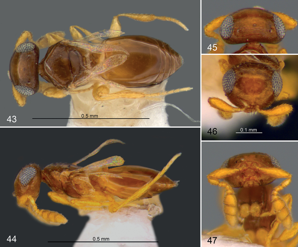
Morphological terms used with abbreviations in parentheses, cross-referenced to an ontological (formal) definition (Hymenoptera Anatomy Ontology; URI = Uniform Resource Identifier) and the figures where these structures are emphasized.
| Terms | definition | URIs | Fig. |
|---|---|---|---|
| HEAD | |||
| antennomere A1, .... A10 | The anatomical structure that is delimited by the proximal and distal margins of the antennal sclerite. | http://purl.obolibrary.org/obo/HAO_0000107 | 1 |
| clypeus (cly) | The area that corresponds to the site of origin of the clypeo-epipharyngeal muscle. | http://purl.obolibrary.org/obo/HAO_0000212 | 2–3 |
| malar sulcus (ms) | The sulcus that extends between the ventral margin of the compound eye and the base of the mandible. | http://purl.obolibrary.org/obo/HAO_0000504 | 4 |
| occiput (occ) | The area that is concave and surrounds the postocciput. | http://purl.obolibrary.org/obo/HAO_0000658 | 8 |
| OD | The diameter of the ocellus. | http://purl.obolibrary.org/obo/HAO_0002107 | 5 |
| ocular ocellar line (OOL) | The anatomical line that is shortest and connects the compound eye and the lateral ocellus. | http://purl.obolibrary.org/obo/HAO_0000662 | 5 |
| paraocellar depressions (paro) | The depressions that flank the lateral margins of the lateral ocelli. |
|
5 |
| preocellar depression (preo) | The depression that flanks the anterior margin of the anterior ocellus. |
|
5 |
| epitorular carina (sc) | The carina that dorsally surrounds the antennal foramen. |
|
2–3 |
| sensillar formula (ps) | Distribution of papillary sensilla (ps) on the ventral clavomeres of the female. |
|
1 |
| torulus (tor) | The foramen that is located on the head in which the radicle is positioned. | http://purl.obolibrary.org/obo/HAO_0001022 | 3 |
| ventral (inner) lamella on A1 (vl) | (Semi)transparent sharp edge on the ventral side of the A1, usually on the apex, but sometimes on the entire length of A1, housing the A2 or A2-A6. | Modified after |
1 |
| MESOSOMA | |||
| antero-admedian line (aadl) | The signum that is submedian and located on the anterior margin of the mesoscutum and corresponds to the site of origin of the longitudinal flight muscle | http://purl.obolibrary.org/obo/HAO_0000128 | 12 |
| axilloaxillular carina (aaxc) | Carina that connects the axillar carina to the axillular carina. Can be regarded as an extension of the axillar carina. | Present study. | 6–8 |
| axillular carina (axc) | The axillular line that is a carina. | http://purl.obolibrary.org/obo/HAO_0000161 | 9–11 |
| transverse pronotal sulcus (cps) | The sulcus that corresponds to the anteromedian pronotal ridge. | http://purl.obolibrary.org/obo/HAO_0001032 | 13 |
| dorsal axillar area (daa) | The area that is located medially on the axilla and is delimited laterally by the axillar carina and posteromedially by scutoscutellar sulcus. | http://purl.obolibrary.org/obo/HAO_0000252 | 14 |
| dorsal metapleural area (dma) | The area that is delimited posterodorsally by the metapleural carina and anteroventrally by the metapleural sulcus. | http://purl.obolibrary.org/obo/HAO_0000261 | 15 |
| mesofemoral depression (fd) | The scrobe that is located on the mesopleuron into which the mesofemur fits when pressed against the mesosoma. | http://purl.obolibrary.org/obo/HAO_0000326 | 16 |
| foamy structure (fs) | Foamy structures are extensions of cuticle that usually emanate from carinae on the propodeum and metapleuron but may also occur on T1 and S1. |
|
15, 17 |
| internotaular area (ina) | The area on the mesoscutum that is delimited laterally by notauli |
|
18 |
| lateral pronotal area (lpa) | The area of the pronotum that is lateral and delimited medially by the epomial carina. | http://purl.obolibrary.org/obo/HAO_0000483 | 19 |
| lateral propodeal carina (lpc) | The carina that is oblique and arises submedially from the anterior margin of the metapectal-propodeal complex and extends to the posterior propodeal projection. | http://purl.obolibrary.org/obo/HAO_0000486 | 21–23 |
| marginal setae of fore wing | The row of setae that is located along the margin of the wing blade in the same plane as the wing blade. | http://purl.obolibrary.org/obo/HAO_0000511 | |
| mesocutellum (mes) | The scutellum that is located on the mesonotum. | http://purl.obolibrary.org/obo/HAO_0000574 | 20 |
| mesopleural carina (mplc – red arrow) | The carina that crosses the mesopleuron and limits ventrally the mesofemoral depression. | http://purl.obolibrary.org/obo/HAO_0000559 | 24 |
| mesopleuron (mpl – marked with red dots) | The area that is located lateral of the mesodiscrimen. | http://purl.obolibrary.org/obo/HAO_0000566 | 24 |
| mesoscutum | The scutum that is located on the mesonotum. | http://purl.obolibrary.org/obo/HAO_0000575 | |
| metapleural carina (mtpc) | The carina that delimits the metapleuron dorsally from the propodeum, extends from just ventral of the metapleural arm to the metacoxal articulation and passes anteroventral to the propodeal spiracle. | http://purl.obolibrary.org/obo/HAO_0000609 | 25, 50 |
| metapleural sulcus (mtps – red arrow) | The line that corresponds with the metapleural ridge. | http://purl.obolibrary.org/obo/HAO_0000614 | 26 |
| metapleuron (mtp – marked with red dots) | The area of the metapectal-propodeal complex that is located laterally of the metadiscrimen. | http://purl.obolibrary.org/obo/HAO_0000621 | 26 |
| metascutellar carina (mtsc) | The carina that delimits laterally the metascutellum. | http://purl.obolibrary.org/obo/HAO_0000624 | 27 |
| metascutellum (mts) | The area that is located posteromedially on the metanotum, is delimited laterally by the metanotal trough and corresponds to the reservoir of the dorsal vessel. | http://purl.obolibrary.org/obo/HAO_0000625 | 27 |
| metasomal depression (metd) | The acetabulum that is concave, surrounds the nucha and accommodates the base of the metasoma. | http://purl.obolibrary.org/obo/HAO_0000627 | 28–29 |
| notauli (nt) | The line that extends submedially along the mesoscutum and corresponds to the median border of the site of origin of the first mesopleuro-mesonotal muscle. | http://purl.obolibrary.org/obo/HAO_0000647 | 22 |
| parapsidal lines | The signum that is located between the notaulus and the parascutal carina and corresponds to the site of origin of the dorsoventral indirect flight muscle. | http://purl.obolibrary.org/obo/HAO_0000694 | |
| plica (pl) | The carina that arises from the anterior margin of the abdominal tergum 1 medially of the propodeal spiracle extends to the posterior propodeal projection. | http://purl.obolibrary.org/obo/HAO_0000735 | 30 |
| posterior mesoscutellar sulcus (pms) | The line that extends along the posterior margin of the mesoscutellum and corresponds to the posterior mesoscutellar ridge. | http://purl.obolibrary.org/obo/HAO_0000757 | 31 |
| prespecular sulcus | The sulcus that delimits anteriorly the speculum and corresponds to the anterior margin of the speculum. | http://purl.obolibrary.org/obo/HAO_0000816 | 42 |
| pronotum (pr) | The notum that is located in the prothorax. | http://purl.obolibrary.org/obo/HAO_0000853 | 32 |
| scuto-scutellar sulcus (sss) | The sulcus that extends along the scutoscutellar suture. | http://purl.obolibrary.org/obo/HAO_0000919 | 33 |
| transepisternal line (tspl) | The line that is longitudinal, extends ventrolaterally on the mesopleuron and corresponds with the site of origin of the second and third mesopleuro-third axillary sclerite of fore wing muscle and the second mesopleuro-mesonotal muscle. | http://purl.obolibrary.org/obo/HAO_0001205 | 34 |
| transscutal articulation (tsa) | The line of separation that extends along the transscutal line. | http://purl.obolibrary.org/obo/HAO_0001204 | 35 |
| METASOMA | |||
| metasomal tergite 1, 2, ... n. (T1–Tn) | The abdominal tergum that is located in the metasoma. | http://purl.obolibrary.org/obo/HAO_0001349 | 36–37 |
| anterior pits of T2 (apT2) | Paired, oval or circular depressions situated anterolaterally on T2, often filled with dense pilosity. | https://doi.org/10.4039/entm121147fv | 36–37 |
The terminology of surface sculpturing is from

Results
Here, we follow the generic concept of Fidiobia presented in
Males of many species of Fidiobia are unknown, rare, or morphologically similar to their female conspecifics. For this reason, we present a key to females; however, given the similarity between males and females of many species, the identification of males may be possible using this key.
Key to Palearctic Fidiobia (females)
| 1 | Antenna 10-merous (Figs |
2 |
| – | Antenna 9-merous (Figs |
7 |
| 2 | Notauli present, incised; junction of T1 and T2 covered by a transverse row of long, strong setae (Fig. |
3 |
| – | Notauli absent (Figs |
5 |
| 3 | Mesoscutellum with reticulate-rugose to longitudinally strigose microsculpture, smooth anteromedially (Figs |
F. striatitergitis (Szabó, 1962) |
| – | Mesoscutellum smooth throughout; T2 smooth; lateral propodeal carina with foamy structures; metasomal depression without median carina; metapleural carina prolonged posterodorsally into a very small tooth | 4 |
| 4 | OOL 5× diameter of lateral ocellus (Figs |
F. nipponica sp. nov. |
| – | OOL at most 3× diameter of posterior ocellus (Fig. |
F. vladlubomiri sp. nov. |
| 5 | Body flattened; marginal setae of fore wing long (Fig. |
F. hispanica Popovici & Buhl, 2010 |
| – | Body not distinctly flattened; marginal setae of fore wing short; posterior side of hind coxa with one or two setose furrows; transepisternal line present; prespecular sulcus absent | 6 |
| 6 | Malar sulcus absent (Fig. |
F. longiclava sp. nov. |
| – | Malar sulcus present (Fig. |
F. tripotini sp. nov. |
| 7 | Notauli present, incised | 8 |
| – | Notauli absent | 23 |
| 8 | Sculpture of frons areolate-rugulose (Figs |
9 |
| – | Sculpture of frons reticulate-coriaceous or alutaceous (Figs |
15 |
| 9 | A1 strongly widened with lamella well developed along the entire ventral margin (Figs |
F. gallica sp. nov. |
| – | A1 moderately widened with lamella present only in the apical third (Figs |
10 |
| 10 | Brachypterous (Figs |
11 |
| – | Macropterous (Figs |
13 |
| 11 | Apex of fore wing tapering to a point (Fig. |
F. pronotata Szabó, 1958 |
| – | Apex of fore wing rounded (Figs |
12 |
| 12 | Apex of fore wing not reaching the middle of T2 (Fig. |
F. pronotatoides sp. nov. |
| – | Apex of fore wing surpassing the middle of T2 (Fig. |
F. rugosifrons Crawford, 1916 |
| 13 | Area between notauli entirely sculptured (Figs |
F. rugosifrons Crawford, 1916 |
| – | Area between notauli smooth at least posteriorly (Figs |
14 |
| 14 | Area between notauli entirely smooth (Figs |
F. roatai sp. nov. |
| – | Area between notauli smooth in posterior half (Figs |
F. rugosifronsoides sp. nov. |
| 15 | Brachypterous species; fore wing reduced, hardly visible (Fig. |
F. hofferi Kozlov, 1978 |
| – | Macropterous species, fore wings extending to or surpassing apex of metasoma (Figs |
6 |
| 16 | Metascutellum not visible in dorsal view, covered by posterior margin of mesoscutellum (Figs |
17 |
| – | Metascutellum visible in dorsal view as a narrow strip bordered by metascutellar carinae (Figs |
8 |
| 17 | Marginal setae of fore wings short (Fig. |
F. brevinotaula Veenakumari et al., 2018 |
| – | Marginal setae of fore wings long (Fig. |
F. insoonae sp. nov. |
| 18 | Epitorular carina present on frons | 19 |
| – | Epitorular carina absent on frons | 21 |
| 19 | Metapleuron with posteroventral third entirely covered with short, dense, white setae (Fig. |
F. communis sp. nov. |
| – | Metapleuron with posteroventral third not entirely covered by setae (Figs |
20 |
| 20 | Metapleuron with a line of stout setae along the dorsal and posterior margins; fore wing dark medially; OOL equal to or less than OD | F. vanharteni Buhl, 2010 |
| – | Metapleuron with only sparse setae, not arranged in a continuous line; fore wings uniformly hyaline; OOL equal to about 2 OD | F. hofferi Kozlov, 1978 |
| 21 | Fore wings with long, visible marginal fringe; T1 with three pairs of sublateral setae (Fig. |
F. bohemica sp. nov. |
| – | Fore wings with hardly visible marginal fringe; T1 with two pairs of sublateral setae; metapleural carina with a very narrow crease of foamy structure; metapleural epicoxal area without a flange of foamy structure over the base of hind coxa | 22 |
| 22 | T2 square or nearly so, at least 4 times as long as T1; notauli parallel; dorsal mesopleuron with numerous delicate, transverse and dense striae; lateral pronotal area sculptured in dorsal two thirds; tibia and scape dark brown | F. platystasioides sp. nov. |
| – | T2 transverse, at most 3 times as long as T1; notauli diverging anteriorly; dorsal mesopleuron with two transverse striae bordering a smooth space; lateral pronotal area sculptured in dorsal third; tibia and scape yellow | F. lisenchiae sp. nov. |
| 23 | Transscutal articulation incomplete (Fig. |
F. sashai sp. nov. |
| – | Transscutal articulation complete; wings macropterous or brachypterous, extending posteriorly beyond propodeum | 4 |
| 24 | Lateral propodeal carinae not connected by a transverse carina (Figs |
25 |
| – | Lateral propodeal carinae sometimes connected by a transverse carina (Figs |
26 |
| 25 | Body flat, strongly depressed dorsoventrally; width of mesosoma at least 2.7 times its height; metascutellum not visible dorsally, covered by posterior margin of mesoscutellum; mesopleuron without large circular depression; transepisternal line complete (Fig. |
F. synergorum (Kieffer, 1921) |
| – | Body not depressed dorsoventrally; width and height of mesosoma nearly equal; metascutellum visible dorsally; mesopleuron with a large circular depression (Fig. |
F. hirta sp. nov. |
| 26 | Transepisternal line present (Fig. |
F. filicornis Buhl, 2014 |
| – | Transepisternal line absent; anterior pits of T2 ovate, distinct separated; median carina between lateral propodeal carinae variable | 27 |
| 27 | T2 distinctly longer than wide (Fig. |
28 |
| – | T2 about as long as wide (Fig. |
29 |
| 28 | Apex of fore wing not extending beyond the middle of T2; ventral third of mesopleuron without longitudinal striae; metapleural sulcus present; metascutellum visible dorsally | F. brevialis sp. nov. |
| – | Apex of fore wings surpassing end of metasoma; ventral third of mesopleuron longitudinally striate; metapleural sulcus absent; metascutellum not visible dorsally, covered by posterior margin of mesoscutellum | F. flaviabdominalis Veenakumari et al., 2018 |
| 29 | Fore wing with long marginal fringe (Figs |
F. polita Buhl, 1998 |
| – | Fore wing with short marginal fringe (Fig. |
F. politoides sp. nov. |
Species descriptions
Fidiobia bohemica , sp. nov.
Description
Female.
Body length: 0.7 mm. Colour of body: melanic (Figs
Head
(Fig.

Mesosoma
(Figs
Metasoma. Posterior of T2 some or all tergites may be retracted under T2. Shape of T1: trapezoidal. Colour of T1: brown. Lateral setae of T1: 3 pairs. Colour of T2: brown. Shape of T2: longer than wide. Anterior pits of T2: distinctly separated. Sculpture of T2, lateral to anterior pits of T2: absent. Colour of T3 –T5: the same as T2.
Male. unknown.
Etymology
Named after the country where the type material was collected. Noun in apposition.
Material examined
2♀. Czech Republic: Holotype 1♀, Orlické Hory, Trčkov, Bukačka res., 50.336°N, 16.372°E, 28.vi–18.vii.1994, leg. Macek J. (MT) (
Distribution
Czech Republic (Fig.
Diagnosis
Fidiobia bohemica is close to F. communis and F. hofferi because of the presence of notauli, the visible metascutellum and the reticulate-coriaceous to alutaceous sculpture of the frons. Fidiobia bohemica differs from these species by the presence of three pairs of sublateral setae on T1 (only two in F. communis and F. hofferi) and the absence of epitorular carinae on the frons (present in F. communis and F. hofferi).
Fidiobia brevialis , sp. nov.
Description
Female. Body length: 0.8 mm. Colour of body: xanthic (Figs
Head
(Figs
Mesosoma
(Figs
Metasoma
(Figs
Male. unknown.
Etymology
The species name is derived from Latin words “brevis” and “alis”, meaning “short wings”.
Material examined
2♀. Japan: Holotype 1♀, Hokkaido Tomuraushi area, 43.45°N, 142.91°E, 13.viii.1996, leg. Masner L. (SS) (
Distribution
Japan (Fig.
Biology
unknown.
Diagnosis
Fidiobia brevialis and F. sashai are the only Palearctic species of the genus that are brachypterous and lack notauli. These species can be separated by the length of the fore wings (hardly longer than the tegula in F. sashai and surpassing the middle of T2 in F. brevialis) and the length of the transscutal articulation (incomplete in F. sashai and complete in F. brevialis).
Fidiobia brevinotaula
Fidiobia brevinotaula Veenakumari, Popovici & Buhl, 2018: 557.
Description
Female (Figs
Head
(Figs

Mesosoma
(Figs
Metasoma
(Fig.
Male. unknown.
Material examined
5♀. Russia: 1♀, Primorsky Krai, Ussuriysk District, Gornotayozhnoye, 44.1°N, 132.41°E, 4–10.viii.1999, leg. Michailovskaya MV. (YPT) (
South Korea: 1♀, Gyeongsan-si, Daehak-ro 280, Yeungnam University, 35.82119°N, 128.7634°E, 14.viii.2016, Fusu L. (YPT) (OPPC0073).
Distribution
India (Veenakumari et al. 2018), Russia, South Korea (Fig.
Diagnosis
Fidiobia brevinotaula is a distinct species based on the abbreviated notauli; the transaxillar carina and horizontal part of the dorsal axillar area that are not visible; the presence of foamy structures on the lateral propodeal carinae; the long, strong, white, dense setae on the metapleuron; and the minute size of specimens. It is close habitually to F. insoonae, but these species can be separated by the marginal setae of fore wings (short in F. brevinotaula and long in F. insoonae) and by the setation of metapleuron (there are long, strong, dense setae in F. brevinotaula and short, tiny, sparse setae in F. insoonae).
Fidiobia communis , sp. nov.
Description
Female. Body length: 0.8–0.9 mm. Colour of body: melanic (Figs
Head
(Fig.

Mesosoma
(Figs
Metasoma. Posterior of T2 some or all tergites may be retracted under T2. Shape of T1: trapezoidal. Colour of T1: brown. Lateral setae of T1: 2 pairs. Colour of T2: brown. Shape of T2: longer than wide. Anterior pits of T2: distinctly separated. Sculpture of T2, lateral to anterior pits of T2: absent. Colour of T3–T5: the same as T2.
Male (Fig.
Etymology
This species is named “communis” because of the absence of any peculiar or striking characters.
Material examined
6♀ and 1♂. Romania: Holotype 1♀, Suceava, Călimani Mts., road of Maria Teresa, 47.12346°N, 25.20249°E, 13–20.vii.2012, leg. Popovici O. (SS) (OPPC0577).
Paratypes
: Czech Republic: 1♀, Bohemia, Celákovice Lipovka Res., 50.177°N, 14.759°E, 2–19.vi.1994, leg. Macek J. (MT) (
Estonia: 2♀, 1.5 km NE Sööru, 58.66111°N, 26.88531°E, 4–11.vii.2011, leg. Soon V. (SN) (OPPC0664, 0665).
Romania: 1♂, Suceava, Călimani Mts., road of Maria Teresa, 47.12346°N, 25.20249°E, 13–20.vii.2012, leg. Popovici O. (SS) (OPPC0578).
Ukraine: 1♀, Transcarpathia, Svydovets, 2–3 km NW of Kvasy, 48.15247°N, 24.26621°E, 7.v–5.vi.2014, leg. Varga O. (TT) (OPPC0230); 1♀, Transcarpathia, Svydovets, 2–3 km NW of Kvasy, 48.15247°N, 24.26621°E, 5–29.vi.2014, leg. Varga O. (TT) (OPPC0146).
Distribution
Czech Republic, Estonia, Romania, Ukraine (Fig.
Diagnosis
Fidiobia communis is close to F. hofferi because of its general habitus, the metascutellum that is visible in dorsal view and the presence of epitorular carinae. These two species differ mainly by the sculpture of the dorsal mesopleuron (reduced in F. hofferi and extending to the middle of the mesopleuron in F. communis), setation of the ventral metapleural area (few, sparse setae in F. hofferi and dense, long setae in F. communis) and the length of the marginal setae on the fore wings (very short, hardly visible in F. hofferi and clearly visible in F. communis).
Fidiobia filicornis
Fidiobia filicornis Buhl, 2014: 74.
Description
Female. Body length: 0.7–08 mm. Colour of body: bicoloured, head and mesosoma dark brown to brown, metasoma brown to reddish brown with T1 and the apex of T6 lighter (Figs
Head
(Figs

Fidiobia filicornis: 61 female, habitus, dorsal view (OPPC0074) 62 female, habitus, lateral view 63 male, habitus, dorsal view (OPPC0045) 64 antenna (♀) (OPPC0517) 65 antenna (♂) (OPPC0818) 66 head and mesosoma, dorsal view 67 head and mesosoma, lateral view 68a wings (OPPC0517) 68b WIP.
Mesosoma
(Figs
Metasoma
(Fig.
Male (Figs
Material examined
86♀ and 39♂. Togo: Holotype ♂, (Figs
China: 1♀ and 2♂, Beijing Prov., Mentougo 39.987°N, 115.5246°E, dry meadow, 28.vii.2002, leg. Melika G. (

South Korea: 2♀, Jirisan, Hamyang-gun, Macheon-myon, Samjeong-li, 35.3486°N, 127.6392°E, 24.viii–15.ix.2003, leg. Tripotin P. (MT) (
Distribution
Togo (
Biology
unknown.
Diagnosis
Fidiobia filicornis is the only known Palearctic species with 9-merous antenna in the female and 10-merous antenna in the male. As is typical for Fidiobia, the female antenna is clavate and the male antenna is clubbed, but in the male of F. filicornis the antenna is almost filiform as in F. longiclava or F. vladlubomiri (both species with 10-merous antenna in male and female). Another distinctive character among the Palearctic species with 9-merous antennae is the presence of the transepisternal line, which is narrow, deeply incised, transverse and nearly complete in F. filicornis. This species is not known from the Oriental region (Veenakumari et al. 2018), but a new species, Fidiobia setosa was recently described from India and is considered a close relative of F. filicornis. These two species can be easily separated because of the presence of a hyperoccipital carina and 10-merous antennae in F. setosa.
Comments
Fidiobia filicornis was described from the Afrotropical region (Togo) by
Fidiobia flaviabdominalis
Fidiobia flaviabdominalis Veenakumari, Popovici & Buhl, 2018: 556, 568.
Description
Female. Body length: 0.5 mm. Colour of body: xanthic, head and mesosoma brown, metasoma light brown to yellow (Figs
Head
(Figs

Mesosoma
(Figs
Metasoma
(Fig.
Material examined
36♀ and 1♂. Japan: 2♀, Kyushu, Fukuoka Mt. Hiko, 33.1259°N, 130.7876°E, 21–29.vii.1989, leg. Takeno K. and Sharkey M. (MT) (
South Korea: 25♀, Jeollabuk-do, Buan-gun Samae-myeon Yuyu village, 35.4191°N, 127.2755°E, 5.vii–14.viii.2007, leg. Tripotin P. (MT) (OPPC0785, 0786, 0787, 0788, 0793, 0792, 0789, 0784, 0790, 0791, 0805, 0809, 0810, 0811, 0806, 0405, 0406, 0647, 0783, 0807, 0404, 0808, 0782, 0477, 0369); 1♀, Jeollabuk-do, Buan-gun Samae-myeon, Yuyu village, 35.4191°N, 127.2755°E, 21.iv–27.v.2007, leg. Tripotin P. (MT) (OPPC0418); 1♀, Chungnam, Daejeon-si, Wadong, 36.3601°N, 127.2345°E, 20.v–19.vi.2007, leg. Tripotin P. (MT) (OPPC485); 2♀, Chungnam, Daejeon-si, Wadong, 36.3601°N, 127.2345°E, 19.vi–24.vii.2007, leg. Tripotin P. (MT) (OPPC0056 – no head, 0054); 1♀, Chungnam, Daejeon-si, Wadong, 36.3601°N, 127.2345°E, 21.viii–25.ix.2007, leg. Tripotin P. (MT) (OPPC0545); 1♀, Chungnam, Daejeon-si, Wadong, 36.3601°N, 127.2345°E, 25.ix–17.xi.2007, leg. Tripotin P. (MT) (OPPC0542); 1♀, Chungbuk, Okcheon-gun Dongi-myeon, Soesan-li, 36.2764°N, 127.6131°E, 8–23.vii.2004, leg. Tripotin P. (MT) (OPPC0728); 2♀, Jirisan, Hamyang, Songjeon-li, Munsu-sa, 35.41232°N, 127.7303°E, 28.vii–16.viii.2004, leg. Tripotin P. (MT) (OPPC0499, 0500).
Distribution
India (Veenakumari et al. 2018), Japan, South Korea (Fig.
Biology
unknown.
Diagnosis
Fidiobia flaviabdominalis is superficially similar in size and general habitus to F. insoonae, F. polita and F. politoides. It differs from F. insoonae mainly by of the absence of notauli (present in F. insoonae) and to F. polita and F. politoides because of the length of T2 (T2 is longer than wide in F. flaviabdominalis and wider than long in F. polita and F. politoides).
Comments
Fidiobia flaviabdominalis is one of the smallest species of the genus in the Palearctic region. It is peculiar among Palearctic Fidiobia because of its reduced size and the light color. Our specimens differ from the original description by the presence of longitudinal striae on the lower third of the mesopleuron and A4 longer than A3 in females.
Fidiobia gallica , sp. nov.
Description
Female. Body length: 1.1 mm. Colour of body: bicoloured, head and mesosoma black, metasoma brown with T1 lighter (T1 light brown to reddish) (Figs
Head
(Figs

Mesosoma
(Figs
Metasoma
(Figs
Male. unknown.
Etymology
This species is named “gallica”, meaning “French”, for the country where the specimen was collected. This species was named after the ancient name of France.
Material examined
1♀. France: Holotype 1♀, Montpellier, 43.73°N, 3.74°E, 12–18.vii.1981, leg. Vayssières JF. (
Distribution
France (Fig.
Biology
unknown.
Diagnosis
Fidiobia gallica is one of the most peculiar species of the genus because of the lamellate scape, elongate A3, reticulate pattern on the disc of the fore wing and a narrow metasomal depression (width of metasomal depression is less than the length of the lateral propodeal carina) bordered by lateral propodeal carinae that are nearly parallel and are elevated posteriorly. The combination of these four characters differentiates this species from the remainder of the Palearctic fauna.
Comments
The development of the ventral lamella of A1 is found in other Palearctic platygastrids, including Iphitrachelus Walker and Amblyaspis Förster. In other regions, this can be found in Sacespalus Kieffer, Platygastoides Dodd, Plutomerus Masner and Huggert, and Pulchrisolia Szabó. The reticulate fore wing can also be found an undescribed species from Madagascar (Z. Lahey, unpublished data).
Fidiobia hirta , sp. nov.
Description
Female. Body length: 1.1 mm. Colour of body: melanic (Figs
Head
(Fig.

Mesosoma
(Figs
Metasoma
(Fig.
Male. unknown.
Etymology
This species is named for the Latin term for hairy, “hirta”.
Material examined
5♀. Russia: Holotype 1♀, Primorsky Krai, Ussuriysk District, Gornotayozhnoye, 44.1000°N, 132.4167°E, 4–10.viii.1999, leg. Michailovskaya M.V. (YPT) (
Paratypes : South Korea: 2♀, Gangwon-do, Chuncheon Nam-myeon, Hudong-ri, in forest, 34.6422°N, 127.6285°E, 25.v–14.vi.2003, leg Tripotin P. (MT); 1♀, Gangwon-do, Chuncheon Nam-myeon, Magog-li, Hongchen river, 37.72977°N, 127.5765°E, 25.v–14.vi.2003, leg. Tripotin P. (MT) (OPPC0067), 1♀, Gangwon-do, Chuncheon Nam-myeon, Magog-li, Hongchen river, 37.72977°N, 127.5765°E, 17.viii–5.ix.2003, leg. Tripotin P. (MT) (OPPC0069).
Distribution
This species was encountered only in Far East Russia and South Korea (Fig.
Biology
unknown.
Diagnosis
Fidiobia hirta differs from other species in the genus because the body is not flattened dorsoventrally, the mesoscutum and mesoscutellum are convex in lateral view, the metasomal depression is large, the lateral propodeal carinae diverge posteriorly, and T3 is at least as long as its maximum width.
Fidiobia hispanica
Fidiobia hispanica
Popovici & Buhl, 2010: 1149;
Description
Female. Body length: 0.7–0.9 mm. Colour of body: melanic (Fig.
Head
(Figs

Mesosoma
(Fig.
Metasoma
(Fig.
Male (Fig.
Material examined
22♀ and 3♂. Spain: Holotype (Fig.

Non-type material
England: 16♀ and 3♂, London, Greenwich, Vanbrugh Pits, reared from a batch of beetle eggs in vacated Andricus lignicola (Hartig, 1840) gall on Quercus robur Linnaeus, 1753, (gall collected 17.i.2010, Notton D.G.) (BMNH); 5♀, London, Greenwich, Vanbrugh Pits, TQ397771 (51.4758°N, 0.0111°E), reared from a batch of beetle eggs in a vacated cell of Synergus umbraculus (Olivier, 1791) in an old Andricus kollari (Hartig, 1843) gall on Quercus robur, (gall collected 14.iii.2010, Notton D.G.) (BMNH).
Distribution
Spain, Ireland, England (
Biology
Diagnosis
The small size and delicate exoskeleton of F. hispanica make this species unmistakable among the Palearctic species with 10-merous antennae. The habitus is somewhat similar to that of F. synergorum and these species have been previously confused (
Fidiobia hofferi
Fidiobia hofferi
Kozlov, 1978: 656;
Description
Female. Body length: 0.5–0.6 mm. Colour of body: melanic (Figs
Head. Colour of head: light brown. Sculpture of head: alutaceous. Sculpture of occiput: transverse alutaceous. Ocellar prominence: absent. Preocellar depression: absent. Paraocellar depressions: absent. OOL / ocellar diameter: OOL around 2 times ocellar diameter. Orientation of lower half of inner orbits: visibly divergent. Sculpture of frons immediately anterior to ocellus: alutaceous. Sculpture of frons immediately dorsal to toruli: the same as the rest of frons, but smoother. Epitorular carina: present. Distance between toruli: equal to the transverse diameter of torulus. Setation of clypeus: two setae. Malar sulcus: absent. Antenna (Figs

Fidiobia hofferi: 103a fully winged specimen, habitus, dorsal view (OPPC0635) 103b antenna in fully winged specimen (♀) (OPPC0638) 104a brachypterous specimen, habitus, dorsal view (OPPC0826) 104b antenna in brachypterous specimen (♀) 105 specimen with extremely brachiptery (OPPC0823) 106 brachypterous specimen, lateral view 107a fully developed wings 107b WIP in fully developed wings 108a brachypterous wings (OPPC0656) 108b WIP in brachypterous wings..
Mesosoma. Colour of mesosoma: brown. Mesosoma: weakly compressed dorsoventrally. Pronotum in dorsal view: narrow, collarlike. Transverse pronotal sulcus: present as a narrow groove along anterior rim of pronotum. Posteroventral end of transverse pronotal sulcus: not dilated. Lateral pronotal area: sculptured only on the dorsal half. Antero-admedian line: absent. Mesoscutum: weakly convex. Parapsidal lines: absent. Sculpture of internotaular area: absent. Notauli: present, incised. Shape of notauli: dilated posteriorly and acute anteriorly. Outer edge of notauli: almost colliniar with axillular carina. Orientation of inner edge of notauli: converging posteriorly. Length of notauli: at most 0.3 times as long as length of mesoscutellum, measured along midline. Length of notaulus / maximum width of notaulus: 3–4 times as long as wide. Distance between notauli: greater than the broadest part of notaulus. Transscutal articulation: complete. Scuto-scutellar sulcus: absent. Fovea on scuto-scutellar sulcus: NA. Mesoscutellum: weakly convex. Shape of mesoscutellum: subrectangular. Axillular carina: posterior apex of axillular carinae touching the posterior edge of mesoscutellum. Axilloaxillular carina: present. Sculpture of mesoscutellum: absent. Posterior mesoscutellar sulcus: absent. Metascutellum: entirely visible. Metascutellar carina: present. Width of metasomal depression: greater than the length of lateral propodeal carina. Median carina between lateral propodeal carinae: absent. Transverse carina between lateral propodeal carinae: present. Foamy structure on transverse carina between lateral propodeal carinae: present. Foamy structure on metasomal depression: absent. Lateral propodeal carinae: parallel. Foamy structure on lateral propodeal carina: present only on the posterior half of the vertical part. Plica: not visible. Posterior end of plica: NA. Foamy structure on plica: NA. Foamy structure on metapleural carina: present on the entire carina. Foamy structure on ventral metapleural area: present. Setation of dorsal metapleural area: few, sparse setae. Setation of ventral metapleural area: few, sparse setae. Longitudinal striation on dorsal mesopleuron: present. Transepisternal line: absent. Mesopleural carina: present. Metapleural sulcus: present, incomplete. Wings (Figs
Metasoma. Posterior of T2 some or all tergites may be retracted under T2. Shape of T1: trapezoidal. Colour of T1: brown. Lateral setae of T1: 2 pairs. Colour of T2: brown. Shape of T2: longer than wide. Anterior pits of T2: distinctly separated. Sculpture of T2, lateral to anterior pits of T2: absent. Colour of T3–T5: the same as T2.
Male. unknown.
Material examined
19♀. Czech Republic: 2♀, (paralectotypes), Moravia, Bzenec, 48.967°N, 17.253°E, 1.vii.1958, leg. Lemarie J., (ex. larva ichneumonid) [OPPC0814 (Figs

Romania: 8♀ (brachypterous) and 8♀ (full winged), Iași, Bârnova forest near Slobozia, 47.01139°N, 27.60306°E, 4.vii.2011, leg. Noyes JS. (SS) (OPPC0660, 0659, 0658, 0657, 0662, 0656, 0826, 0661 and OPPC0635, 0636, 0637, 0655, 0633, 0663, 0638, 0634).
Ukraine: 1♀, Transcarpathia reg., Svydovets, 2–3 km NW of Kvasy, 48.1524°N, 24.2662°E, 5–29.vi.2014, beech forest, leg. Varga O. (TT) (OPPC0823).
Distribution
Finland, Sweden, Iran (
Biology
The host is unknown, but
Diagnosis
This species can be diagnosed by the visible metascutellum and nearly glabrous metapleuron. It is relatively close to F. vanharteni and F. polita based on its general habitus. Fidiobia hofferi is most likely to be confused with F. polita, a species with which it is sympatric. The main difference is the presence of notauli in F. hofferi and the absence of these structures in F. polita. Another difference between these two species is the OOL:OD ratio (OOL is 2 times as long as OD in F. hofferi and OOL is equal to OD in F. polita).
Fidiobia hofferi can be separated from F. vanharteni because the fore wings are uniformly hyaline in F. hofferi and dark medially in F. vanharteni. Also, the OOL is equal to about 2 OD in F. hofferi and the OOL is equal to or less than OD in F. vanharteni. Fidiobia hofferi is a polymorphic species and contains brachypterous females among the Romanian material.
Comments
In specimens from the type series, the median prominence of T1 is smooth and without carinae. In the Romanian material, the median prominence of T1 has two carinae. Also, the specimens from Romania are more gracile than the specimens from the type series. The specimen from Ukraine has the wings more reduced than the brachypterous specimens from Romania, which are about half the length of the notauli, and the medial prominence of T1 with three carinae. The Ukrainian specimen otherwise matches our concept of F. hofferi.
Fidiobia insoonae , sp. nov.
Description
Female. Body length: 0.5 mm. Colour of body: melanic (Figs
Head. Colour of head: brown. Sculpture of head: reticulate-coriaceous. Sculpture of occiput: transverse reticulate coriaceous. Ocellar prominence: absent. Preocellar depression: absent. Paraocellar depressions: absent. OOL / ocellar diameter: OOL equal with ocellar diameter. Orientation of lower half of inner orbits: visibly divergent. Sculpture of frons immediately anterior to ocellus: alutaceous. Sculpture of frons immediately dorsal to toruli: the same with the sculpture from the rest of frons, but more transverse. Epitorular carina: present. Distance between toruli: equal to the transverse diameter of torulus. Setation of clypeus: two setae. Malar sulcus: absent. Antenna (Fig.

Mesosoma
(Figs
Metasoma. Posterior of T2 some or all tergites may be retracted under T2. Shape of T1: trapezoidal. Colour of T1: brown. Lateral setae of T1: 2 pairs. Colour of T2: brown. Shape of T2: longer than wide. Anterior pits of T2: distinctly separated. Sculpture of T2, lateral to anterior pits of T2: absent. Colour of T3–T5: the same as T2.
Male. unknown.
Etymology
This species is named in honor of Insoon Tripotin.
Material examined
8♀. South Korea: Holotype 1♀, Chungnam, Daejeon-si, Wadong, 36.3601°N, 127.2345°E, 19.vi–24.vii.2007, leg. Tripotin P. (MT) (OPPC0058).
Paratypes : South Korea: 1♀, Chungnam, Daejeon-si, Wadong, 36.3601°N, 127.2345°E, 20.v–19.vi.2007, leg. Tripotin P. (MT) (OPPC0064); 1♀, Chungnam, Daejeon-si, Wadong, 36.3601°N, 127.2345°E, 19.vi–24.vii.2007, leg. Tripotin P. (MT) (OPPC0057); 1♀, Chungnam, Daejeon-si, Wadong, 36.3601°N, 127.2345°E, 21.viii–25.ix.2007, leg. Tripotin P. (MT) (OPPC0546); 1♀, Chungnam, Daejeon-si, Wadong, 36.3601°N, 127.2345°E, 25.ix–17.xi.2007, leg. Tripotin P. (MT) (OPPC0544); 1♀, Gangwon-do, Chuncheon Nam-myeon, Magog-li, Hongchen river, 37.72977°N, 127.5765°E, 11.vii–7.viii.2004, leg. Tripotin P. (MT) (OPPC0643); 1♀, Gangwon-do, Chuncheon Nam-myeon, Magog-li, Hongchen river, 37.72977°N, 127.5765°E, 26.ix–31.x.2004, leg. Tripotin P. (MT) (OPPC0816); 1♀, Jirisan, Hamyang, Songjeon-li, Munsu-sa, 35.41232°N, 127.7303°E, 6–27.vi.2004, leg. Tripotin P. (MT) (OPPC0493).
Distribution
South Korea (Fig.
Diagnosis
Fidiobia insoonae is superficially similar to F. polita, F. politoides, F. flaviabdominalis, and F. hofferi because of the almost similar size and the general habitus. It most obviously differs from F. polita, F. politoides, and F. flaviabdominalis (it is sympatric with the latter two) by the presence of notauli. Fidiobia insoonae and F. hofferi are allopatric and differ from each other mainly by the metascutellum, which is covered by the posterior margin of mesoscutellum and not visible in F. insoonae, and because of the setation of the metapleuron is sparse in F. hofferi and dense in F. insoonae. Also, the marginal fringe of the fore wing is short and barely noticable in F. hofferi but it is long in F. insoonae.
Fidiobia lisenchiae , sp. nov.
Description
Female. Body length: 0.7 mm. Colour of body: melanic (Fig.
Head
(Figs

Mesosoma
(Figs
Metasoma
(Fig.
Male. unknown.
Etymology
This species is named after Camelia Lisenchi because of her great support during a collecting trip in Cyprus.
Material examined
1♀. Cyprus: Holotype 1♀, 6 km N of Lemessos, 34.727°N, 33.05°E, 24.v.2009, leg. Popovici O. and Fusu L. (SN) (OPPC0813).
Distribution
Cyprus (Fig.
Diagnosis
Fidiobia lisenchiae is similar to F. platystasioides because of the absence of epitorular carinae, the fore wings with very short marginal setae and the notauli slightly dilated posteriorly. These two species are easily separated because the mesosoma is slightly flattened in F. lisenchiae and visibly flattened in F. platystasioides. Also, T2 is transverse in F. lisenchiae and square or nearly so in F. platystasioides. The difference between these two states of T2 is reflected in the ratio of T2:T1. T2 is at most 3 times as long as T1 in F. lisenchiae and at least 4 times as long as T1 in F. platystasioides. The submarginal vein is shorter in F. lisenchiae than in F. platystasioides, with the apex of the submarginal vein hardly surpassing the posterior edge of the propodeum in F. lisenchiae and surpassing the middle of T1 in F. platystasioides. Other subtle differences between these species are the color of the scape and tibia (yellow in F. lisenchiae and dark brown in F. platystasioides), the sculpture of the dorsal mesopleuron (with few striae and a smooth area in F. lisenchiae and with numerous, dense striae in F. platystasiodes) and in the sculpture of the lateral pronotal area (sculptured only in dorsal third in F. lisenchiae and in dorsal two thirds in F. platystasioides).
Fidiobia longiclava , sp. nov.
Description
Female. Body length: 0.8–1.0 mm. Colour of body: Variable, melanic specimens are brown with hardly lighter T1; xanthic specimens are light brown to yellow with darker head (Figs
Head
(Figs

Mesosoma
(Figs
Metasoma
(Figs
Male (Figs

Etymology
This species is named for the elongate shape of the clavomeres.
Material examined
8♀ and 1♂. South Korea: Holotype 1♀, Jirisan, Hamyang-gun, Macheon-myon, Samjeong-li, 35.3486°N, 127.6392°E, 24.viii–15.ix.2003, leg. Tripotin P. (MT) (
Paratypes
: South Korea, 4♀, Jirisan, Hamyang-gun, Macheon-myon, Samjeong-li, 35.3486°N, 127.6392°E, 24.viii–15.ix.2003, leg. Tripotin P. (MT) (
Distribution
South Korea (Fig.
Biology
unknown.
Diagnosis
The most diagnostic feature is the elongated shape of the clavomeres, which are unique among the Palearctic species of Fidiobia with 10-merous antennae. Non-sexually dimorphic characters that can be used to identify males are the notaular lines that are visible as a change in the setation of the mesoscutum and the nearly straight transepisternal line. The transverse anterior pits of T2 that nearly merge medially is unique among Palearctic Fidiobia that have a 10-merous antenna, lack notauli and have the junction of T1–T2 not covered by a row of setae. The color of the body in this species is highly variable, ranging from almost entirely yellow to completely brown.
Fidiobia nipponica , sp. nov.
Description
Females. Length of body: 1.1 mm. Colour of body: melanic species (Figs
Head
(Figs

Mesosoma
(Figs
Metasoma
(Figs
Male. unknown.
Etymology
This species is named after the country where the type material was collected.
Material examined
1♀. Japan: Holotype 1♀, Tochigi, Hikinuma, Shiobara, 21.viii.1985, leg. Takahaghi K. (TT) (
Distribution
Japan (Fig.
Biology
unknown.
Diagnosis
We consider F. nipponica to be close to F. striatitergitis based on the presence of a metascutellar carina with a tooth, a posterior mesoscutellar sulcus and the very short marginal fringe of the fore wing. Although most of T2 in this species is smooth and shining, some very fine longitudinal striae can be observed laterally, but this sculpture is distinctly different than the extensive striation on T2 that is found in F. striatitergitis. The metasomal depression is completely covered with foamy structures and the large distance between the posterior ocellus and compound eye make this species easy to recognize among the Palearctic species of Fidiobia with 10-merous antennae.
Fidiobia platystasioides , sp. nov.
Description
Female. Body length: 0.7 mm. Colour of body: melanic (Figs

Head
(Figs
Mesosoma
(Figs
Metasoma
(Fig.
Male. unknown.
Etymology
This species is named for its similarity with species of Platystasius Nixon, 1937.
Material examined
1♀. China: Holotype 1♀, Beijing Prov., Mentougo, 39.987°N, 115.5246°E, 28.vii.2002, leg. Melika G. (
Distribution
China (Fig.
Biology
unknown.
Diagnosis
Fidiobia platystasioides is similar to F. lisenchiae because of the absence of epitorular carinae, the fore wings with very short marginal setae and notauli slightly dilated posteriorly. The main differences between these two species is the ratio T2:T1 (in F. platystasioides T2 is at least 4 times as long as T1 and in F. lisenchiae T2 is at most 3 times as long as T1) and the length of the submarginal vein (the apex of the submarginal vein surpassing the middle of T1 in F. platystasioides and hardly surpassing the propodeum in F. lisenchiae). Other subtle differences between these species are the color of the scape and tibia (dark brown in F. platystasioides and yellow in F. lisenchiae), the sculpture of the dorsal mesopleuron (with numerous dense striae in F. platystasiodes and with few striae and a smooth area in F. lisenchiae) and the sculpture of the lateral pronotal area (sculptured in dorsal two thirds in F. platystasioides and only in dorsal third in F. lisenchiae).
Comments
Fidiobia platystasioides is a distinct species because the notauli are slightly dilated posteriorly and almost parallel, and the body is flattened. These characteristics closely resemble those seen in the genus Platystasius Nixon.
Fidiobia polita
Fidiobia polita
Buhl, 1998: 298;
Description
Female. Body length: 0.5–0.6 mm. Colour of body: melanic (Figs

Head
(Figs
Mesosoma
(Figs
Metasoma
(Figs
Male. unknown.
Material examined
14♀. Estonia: 1♀, 1,5 km NE Sööru, 58.65063°N, 26.88531°E, 3–10.vi.2011, leg. Soon V. (OPPC0547).
Greece: 1♀, Kerkini lake Natural Park, Kerkini Mts., 41.28642°N, 23.20147°E, 4.v.2010, leg. Popovici O. and Fusu L. (SN) (OPPC0580).
Hungary: 5♀, Örseg, Nemzeti Park, Lugosy Valley, 46.9°N, 16.45°E, 28.vi.2010, leg. Noyes JS. (SS) (OPPC0817, 0584, 0585, 0587, 0586).
Romania: 1♀, Iași, Ciric, 47.24333°N, 27.57927°E, 20.v.2006, leg. Popovici O. and Moglan I. (SN) (OPPC0798).
Sweden: Holotype ♀, (Figs

Non-type materia
2♀, Småland, Asa, 57.16667°N, 14.78333°E, 5–6.vii.2007, leg. Shevtsova E. (OPPC0738, 0732).
Ukraine: 2♀, Mochary reg., 5 km NE of Bogorodchany, 48.84755°N, 24.59081°E, 16.vi–14.vii.2014, leg. Varga O. (OPPC0055, 0140); 1♀, Mochary reg., 5 km NE of Bogorodchany, 48.84755°N, 24.59081°E, 8–14.vi.2015, leg. Varga O. (OPPC0186).
Distribution
Estonia, Greece, Hungary, Romania, Sweden, Ukraine (Fig.
Biology
unknown.
Diagnosis
Fidiobia polita is distinct among the Palearctic species of this genus with 9-merous antennae and without notauli because T2 is transverse or about as long as wide and the OOL is about as long as an OD (OOL 0.8–1.2 times as long as OD). Of the Palearctic fauna, F. polita is most similar to F. politoides and differs in the length of the fore wing marginal setae (long marginal setae in F. polita and very short marginal setae in F. politoides). According to the studied material these species are allopatric.
In the European fauna, F. polita is similar to F. hofferi but differs by the notauli (which are present in F. hofferi and absent in F. polita) and by the ratio OOL:OD (OOL = 2OD in F. hofferi and OOL = OD in F. polita). Because of the small size of both species, and because in some specimens of F. hofferi the notauli are superficial, the presence of notauli can be difficult to observe. Minor differences can also be observed in the structure of the antenna: A3 is shorter than A4 and the junction between A2 and A3 is narrow in F. polita, but in F. hofferi A3 is almost as long as A4 and the junction between A2 and A3 is large. In F. polita, A5 has the same shape as A4 (globular or moniliform), but in F. hofferi A5 it is more transverse than A4. The study of these characters requires examination of the antenna on a microscopic slide.
Comments
In our material, the specimens from Sweden (type locality) are very similar to the specimen from Estonia. The specimen from Greece is the smallest, relatively weakly sclerotized and, in connection with this, the color of body is lighter than in the rest of the specimens. However, there are no characters to reliably separate it. Also, the Romanian specimen is slightly larger than the rest of the specimens examined.
Fidiobia politoides , sp. nov.
Description
Female. Body length: 0.5 mm. Colour of body: melanic (Figs
Head
(Fig.

Mesosoma
(Figs
Metasoma
(Figs
Male. unknown.
Etymology
This species is named for its similarity to F. polita.
Material examined
2♀. South Korea: Holotype 1♀, Chungnam, Daejeon-si, Wadong, 36.3601°N, 127.2345°E, 24.iv–20.v.2007, leg. Tripotin P. (MT) (OPPC0653).
Paratype : South Korea: 1♀, Gangwon-do, Chuncheon Nam-myeon, Hudong-ri, 34.6422°N, 127.6285°E, 25.v–14.vi.2003, leg Tripotin P. (MT) (OPPC0475).
Distribution
South Korea (Fig.
Biology
unknown.
Diagnosis
Fidiobia politoides is close to F. polita because T2 is transverse or about as long as wide and the OOL is about as long as an OD (OOL 0.8–1.2 times as long as OD) and they differ in the length of the fore wing marginal setae (long marginal setae in F. polita and very short marginal setae in F. politoides). In the structure of antenna, the clava in F. politoides is larger than in F. polita, and A5 and A6 are more transverse in F. politoides than in F. polita. According to the studied material these species are allopatric.
Fidiobia pronotata
Fidiobia pronotata
Szabó, 1958: 459;
Description
Female. Body length: 0.9–1.0 mm. Colour of body: bicoloured, head and mesosoma black, metasoma brown (Figs
Head
(Figs

Mesosoma
(Figs
Metasoma
(Figs
Male. unknown.
Material examined
11♀. France: 1♀, Côte-d’Or, Esbarres, 47.102°N, 5.229°E, 1.ix.1948, leg. Barbier J. (MNHP); 1♀, Côte-d’Or, Esbarres, 47.102°N, 5.229°E, 22.ix.1955, leg. Barbier J. (MNHP); 1♀, Côte-d’Or, Gevrolles, 47.985°N, 4.772°E, 4.ix.1957, leg. Barbier J. (MNHP); 1♀, Côte-d’Or, Esbarres, 47.102°N, 5.229°E, 16.vii.1958, leg. Barbier J. (
Hungary: Holotype: ♀, Pesta, Szentendrei-sziget, 47.643°N, 19.099°E, 2.vii.1957, leg. Szabó JB. (

Romania: 1♀, Iași, Botanical Garden, 47.186°N, 27.5512°E, 17.ix.2003, leg. Popovici O. (sweep net) (OPPC0692); 1♀, Constanța, Vadu, 44.47265°N, 28.8064°E, 26.viii.2004, leg. Popovici O. (sweep net) (OPPC0693); 1♀, Iași, Ciric lake, 47.18778°N, 27.60139°E, 30.vii.2010, leg. Popovici O. (YPT) (OPPC0482); 1♀, Iași, Botanical Garden, 47.1875°N, 27.54889°E, 30.vi.2011, leg. Noyes JS. (SS) (OPPC0755).
Distribution
Germany (
Biology
unknown.
Diagnosis
Fidiobia pronotata can be easily identified by the elongate pronotum, shortened wings and large, non-foveate mesoscutal humeral and suprahumeral sulci. The epomial carina is absent, or very short and weakly indicated. This combination of characters is unique among Palearctic Fidiobia.
Comments
In the original description of this species,
It is a relatively rare species not often collected with sweep nets or Malaise traps.
Fidiobia pronotatoides , sp. nov.
Description
Female. Body length: 0.84 mm. Colour of body: melanic (Figs
Head
(Figs

Mesosoma
(Figs
Metasoma
(Figs
Male. unknown.
Etymology
This species is named for its similarity to F. pronotata.
Material examined
1♀. Romania: Holotype 1♀, Iași, Valea lui David, 47.1939°N, 27.4697°E, 30.v.2018, leg. Popovici O. (SS) (OPPC0001).
Distribution
Romania (Fig.
Biology
unknown.
Diagnosis
There are three brachypterous species of Fidiobia with notauli and areolate-rugulose sculpture on the frons: F. pronotanoides, F. pronotata, and F. rugosifrons. Between these species, brachyptery is always observed in F. pronotata and F. pronotatoides, but it is a rarity for F. rugosifrons.
Fidiobia pronotatoides is very close to F. pronotata, differing by the pronotum of typical length, the narrow mesoscutal humeral and suprahumeral sulci, the well developed epomial carina (longer than half the length of the pronotum measured along midline), fore wings apically rounded (acuminate in F. pronotata), legs light brown with brown coxae (legs entirely yellow in F. pronotata) and a glabrous median prominence of T1 (setose in F. pronotata). Fidiobia pronotatoides can be separated from F. rugosifrons by the length of the fore wing (not reaching the middle of T2), the internotaular sculpture (smooth in posterior half), and the lateral pronotal area (smooth in ventral half).
Fidiobia roatai , sp. nov.
Description
Female. Body length: 0.9–1.0 mm. Colour of body: melanic (Figs
Head
(Figs

Mesosoma
(Figs
Metasoma
(Fig.
Male. unknown.
Material examined
7♀. Romania: Holotype 1♀, Iași, Mârzești, 47.242716°N, 27.471497°E, 19.vi.2016, leg. Popovici O. (SS) (OPPC0827). Paratypes: 2♀, Iași, Mârzești, 47.24417°N, 27.48278°E, 5.vii.2011, leg. Mitroiu M. (SN) (OPPC0567, 0568); 3♀, Iași, Mârzești, 47.242716°N, 27.471497°E, 19.vi.2016, leg. Popovici O. (SS) (OPPC0550, 0540, 0548).
Non-type material
1♀, Iași, Ciric, 26.vi.2006, 47.24333°N, 27.57927°E, leg. Popovici O. (SN) (OPPC0696).
Etymology
This species is named after Dr. Cristian Roată, a well-known surgeon from Iași (Romania).
Distribution
Romania (Fig.
Biology
The host is unknown. The specimens were collected from a typically steppic habitat.
Diagnosis
Fidiobia roatai is distinct among species with an areolate-rugulose frons because of its dark body, absence of sculpture between the notauli, absence of foamy structures on the propodeum, the ratio between A2 and A3 (A2 1.3–1.5 times as long as A3 in F. roatai and 2.4–2.6 times as long as A3 in F. rugosifronsoides), the ratio between A3 and A4 (A3 1.8–2.0 times as long as A4 in F. roatai and 1.2–1.3 times as long as A4 in F. rugosifronsoides) and the rudimentary or absent submarginal vein of fore wing. Also, the fore wings in F. roatai have a peculiar pattern in color and in the distribution of setae. The basal 1/5 of the fore wing is light brown and the setae are absent or punctiform on this area. The apical 3/5 of fore wing is also light brown but covered with short setae. Between basal and apical brown areas of the fore wing there is a lighter almost triangular area. The anterior margin of the fore wings is also peculiar with an expanded costal lobe.
Fidiobia rugosifrons
Fidiobia rugosifrons:
Fidiobia tatrae:
Rosneta phryne:
Fidiobia phryne:
Description
Female. Body length: 0.7–1.0 mm. Colour of body: melanic (Figs
Head. Colour of head: black. Sculpture of head: areolate-rugulose. Sculpture of occiput: areolate-rugulose. Ocellar prominence: absent. Preocellar depression: absent. Paraocellar depressions: absent. OOL / ocellar diameter: OOL equal with ocellar diameter. Orientation of lower half of inner orbits: visibly divergent. Sculpture of frons immediately anterior to ocellus: areolate-rugulose. Sculpture of frons immediately dorsal to toruli: areolate-rugulose. Epitorular carina: absent. Distance between toruli: toruli touch each other. Setation of clypeus: two setae. Malar sulcus: absent. Antenna (Fig.

Mesosoma. Colour of mesosoma: black. Mesosoma: weakly compressed dorsoventrally. Pronotum in dorsal view: narrow, collarlike. Transverse pronotal sulcus: present as a narrow groove along anterior rim of pronotum. Posteroventral end of transverse pronotal sulcus: not dilated. Lateral pronotal area: entirely sculptured. Antero-admedian line: present. Mesoscutum: weakly convex. Parapsidal lines: absent. Sculpture of internotaular area: reticulate rugose. Notauli: present, incised. Shape of notauli: dilated posteriorly and acute anteriorly. Outer edge of notauli: almost collinear with axillular carina. Orientation of inner edge of notauli: converging posteriorly. Length of notauli: half of length of mesoscutum, measured along midline. Length of notaulus / maximum width of notaulus: 2.0–2.9 times as long as wide. Distance between notauli: greater than the broadest part of notaulus. Transscutal articulation: complete. Scuto-scutellar sulcus: absent. Fovea on scuto-scutellar sulcus: NA. Mesoscutellum: weakly convex. Shape of mesoscutellum: subrectangular. Axillular carina: posterior apex of axillular carinae abutting the posterior edge of mesoscutellum. Axilloaxillular carina: present. Sculpture of mesoscutellum: absent. Posterior mesoscutellar sulcus: absent. Metascutellum: visible, partially covered by mesoscutellum. Metascutellar carina: present. Width of metasomal depression: greater than the length of lateral propodeal carina. Median carina between lateral propodeal carinae: absent. Transverse carina between lateral propodeal carinae: present. Foamy structure on transverse carina between lateral propodeal carinae: present. Foamy structure on metasomal depression: absent. Lateral propodeal carinae: parallel. Foamy structure on lateral propodeal carina: present only on the posterior half of the vertical part. Plica: visible. Posterior end of plica: fused with metapleural carina. Foamy structure on plica: present, fused with foamy structure from metapleural carinae. Foamy structure on metapleural carina: present, only posteriorly. Foamy structure on ventral metapleural area: absent. Setation of dorsal metapleural area: dense, long hairs on posterodorsal half. Setation of ventral metapleural area: dense, long hairs on posteroventral half. Longitudinal striation on dorsal mesopleuron: present. Transepisternal line: visible as a ridge on the anteroventral mesopleuron connected with a pit. Mesopleural carina: present. Metapleural sulcus: present, complete. Wings (Fig.
Metasoma. Tergites posterior to T2 may be retracted under T2. Shape of T1: trapezoidal. Colour of T1: dark brown. Lateral setae of T1: 3 pairs. Colour of T2: dark brown. Shape of T2: longer than wide. Anterior pits of T2: distinctly separated. Sculpture of T2, lateral to anterior pits of T2: absent. Colour of T3–T6: the same as T2.
Male. similar to the female, but differs in the structure of the antenna (Fig.
Material examined
29♀ and 3♂. USA: Holotype of F. rugosifrons Crawford (Figs

Belgium: Holotype of Rosneta phryne Debauche (Figs
Hungary: Type of F. tatrae Szelényi (Figs
Non-type material
Hungary: 2♀, Örseg, Nemzeti Park, Lugosy Valley, 46.9°N, 16.45°E, 28.vi.2010, leg. Noyes JS. (SS) (OPPC0582, 0583); 7♀, Vas Co, Köseg, 47.36633°N, 16.52173°E, 26.vi.2010, leg. Hansson C. (SS) (OPPC0707, 0700, 0698, 0708, 0701, 0702, 0699); 1♀, Vas Co, Köseg, 47.36633°N, 16.52173°E, 26.vi.2010, leg. Popovici O. (SN) (OPPC0703); 1♀, Vas Co, Bárkás Lake, 46.86982°N, 16.42605°E, 28.vi.2010, leg. Hansson C. (SS) (OPPC0706).
Estonia: 1♀, 1.5 km NE Sööru, 58.66111°N, 26.88531°E, 21.iv–11.v.2011, leg. Soon V. (SN) (OPPC0590).
France: 1♂, Puy de Dôme, Gergovie Plant., 45.71°N, 3.01°E, 16.vii.1977, leg. de V. Graham MWR (BMNH).
Germany: 2♀, Kiel, leg. Boness M. (BMNH).
Romania: 1♀, Iași, Breazu, 47.2187°N, 27.5270°E, 30.vi.2002, leg. Popovici O. (SN) (OPPC0695); 1♀, Suceava, Todirescu, 47.4455°N, 25.6138°E, 24.vii.2004, leg. Popovici O. and Fusu L. (SN) (OPPC0803); 1♀, Tulcea, Măcin, 45.2358°N, 28.1995°E, 10.vii.2004, leg. Mitroiu M. (SN) (OPPC0694); 1♂, Botoșani, Roma, 1.v.2005, 47.8362°N, 26.5806°E, leg. Popovici M. (SN) (OPPC0691); 1♀, Iași, Botanical Garden, 47.1859°N, 27.5511°E, 21.vi.2005, leg. Popovici O. (SN) (OPPC0812); 1♀, Iași, Bârnova, 46.9938°N, 27.5906°E, 8.vii.2008, leg. Popovici M. (SN) (OPPC0697); 1♀, Iași, Bârnova, 46.9863°N, 27.5855°E, 11.vii.2009, leg. Popovici O. and Popovici M. (SN) (OPPC0490); 1♀, Bacău, Comănești, 46.4288°N, 26.4368°E, 26–31.iv.2013, leg. Pintilioaie A. (SN) (OPPC0576); 1♀, Suceava, Gura Humorului, 47.5563°N, 25.8588°E, 12.v.2013, leg. Bârsan I, (MT) (OPPC0005); 1♀, Tulcea, Periprava, 46.99897°N, 25.94753°E, 8.vii.2015, leg. Popovici O. (SS) (OPPC0829); 1♀ (brachypterous specimen), Suceava, Neagra Șarului, 47.26056°N, 25.35278°E, 3.vii.2011, leg. Noyes JS. (SS) (OPPC0474); 1♀, Iași, Bârnova, 46.9865°N, 27.5839°E, 26.vi.2016, leg. Popovici O. (SS) (OPPC0006); 2♀, Botoșani, Popeni, 47.836832°N, 26.495561°E, 29.vii.2016 leg. Popovici O. (SS) (OPPC007, 0566); 1♂ and 1♀, Harghita, Sovata, 46.569175°N, 25.081698°E, 27.v.2018, leg. Popovici O. (SS) (OPPC0002 and OPPC0003).
Distribution
Asia: Central Altai, Kazakhstan, Central Asia (
Biology
reared from the eggs of Hypera punctata (F) (Coleoptera: Curculionidae) on Triticum sp. (
Diagnosis
Fidiobia rugosifrons is very close to F. rugosifronsoides and F. roatai because of the general habitus and the sculpture of the head, espeacially the frons. Based on this revision, the main characteristics of F. rugosifrons are the totally sculptured internotaular area (unsculptured in F. roatai, or partially sculptured in F. rugosifronsoides), totally sculptured lateral pronotal area (sculptured only on the dorsal half in F. rugosifronsoides and only in the dorsal third in F. roatai) and A3 1.5 times as long as A4 (A3 1.8–2.0 times as long as A4 in F. roatai and 1.2–1.3 times in F. rugosifronsoides).
Comments
The sculptured internotaular area was mentioned by
We located the type of F. tatrae in
Prior to this study, we believe that the name “rugosifrons” was used for a complex of species including F. rugosifrons, F. rugosifronsoides and F. roatai. Although F. rugosifrons was considered as a species with a wide distribution (Fig.
Fidiobia rugosifronsoides , sp. nov.
Description
Female. Body length: 0.9–1.0 mm. Colour of body (Figs

Head
(Figs
Mesosoma
(Figs
Metasoma
(Figs
Male. unknown.
Etymology
This species is named for its similarity to F. rugosifrons.
Material examined
8♀. Finland: Holotype 1♀, Lkor, Sodankylä, Jeesiö, Nurmiharju, 67.508°N, 26.035°E, 11–18.vii.2014, leg. Flinck J. and Aaltio J. (MT) (OOPC0042).
Paratypes : Estonia: 1♀, 1.5 km NE Sööru, 58.66111°N, 26.88531°E, 4–11.vii.2011, leg. Soon V. (SN) (OPPC0593); 2♀, 1.5 km NE Sööru, 58.66111°N, 26.88531°E, 21.vii–13.viii.2011, leg. Soon V. (SN) (OPPC0681, 0652).
Finland: 1♀, Lkor, Sodankylä, Jeesiö, Nurmiharju, 67.508°N, 26.035°E, 11–18.vii.2014, leg. Flinck J. and Aaltio J. (MT) (OOPC0041); 1♀, Lkor, Sodankylä, Jeesiö, Nurmiharju, 67.508°N, 26.035°E, 18–25.vii.2014, leg. Flinck J. and Aaltio J. (MT) (OOPC0040).
Sweden: 1♀, Småland, Asa, 57.16667°N, 14.78333°E, 6.vi.2007, leg. Shevtsova E. (OPPC0731); 1♀, Skåne, Häckeberga, 55.58333°N, 13.41667°E, 5.vii.2006, leg. Hansson C. and Shevtsova E. (OPPC0730).
Non-type material
China: 1♀, Beijing Prov. Mentougo, 39.987°N, 115.5246°E, 28.vii.2002, leg. Melika G. (
South Korea: 8♀, Gangwon-do, Chuncheon Nam-myeon, Magog-li, Hongchen river, 37.72977°N, 127.5765°E, 7.viii–14.ix.2004, leg. Tripotin P. (MT) (OPPC0764, 0741, 0723, 0781, 0749, 0640, 0828, 0487); 1♀, Chungnam, Daejeon-si, Wadong, 36.3601°N, 127.2345°E, 24.iv.–20.v.2007, leg. Tripotin P. (MT) (OPPC0654); 3♀, Chungnam, Daejeon-si, Wadong, 36.3601°N, 127.2345°E, 19.vi–24.vii.2007, leg. Tripotin P. (MT) (OPPC0049, 0050, 0004).
Distribution
Estonia, Finland, Sweden, China, South Korea (Fig.
Biology
unknown.
Diagnosis
Fidiobia rugosifronsoides is close to F. rugosifrons. The main differences between these two species consist of the sculpture of the area between the notauli (smooth in the posterior half in F. rugosifronsoides and totally sculptured in F. rugosifrons), in the ratio of A3 to A4 (A3 1.2 times as long as A4 in F. rugosifronsoides and A3 1.5 times as long as A4 in rugosifrons) and in the sculpture of the lateral pronotal area (entirely sculptured in F. rugosifrons and sculptured only in the dorsal half in F. rugosifronsoides).
Comments
In our material we found this species in Europe in Estonia (here, it is sympatric with F. rugosifrons), Finland and Sweden and in Asia in China and South Korea. Striation below the tegula and longitudinal sculpture below the mesofemoral depression are more evident in specimens from Europe than in the Asian material; the striae of T1 are longer and coarser in the European material; T1 and sometimes the proximal half of T2 is lighter, almost pale in the Asian material and brown in the European material; notauli are broader, and the distance between the medial margin of the notaulus near the transscutal articulation is greater in the European material than in the Asian material.
Fidiobia sashai , sp. nov.
Description
Female. Body length: 0.6 mm. Colour of body: xanthic, brown head and light brown mesosoma and metasoma (Figs
Head
(Figs

Mesosoma. Colour of mesosoma: light brown. Mesosoma: weakly compressed dorsoventrally. Pronotum in dorsal view: cervical pronotal area broader than lateral shoulders. Transverse pronotal sulcus: present as a narrow groove along anterior rim of pronotum. Posteroventral end of transverse pronotal sulcus: not dilated. Lateral pronotal area: sculptured only on the dorsal half. Antero-admedian line: absent. Mesoscutum: flat. Parapsidal lines: absent. Sculpture of internotaular area: absent. Notauli: absent. Shape of notauli: NA. Outer edge of notauli: NA. Orientation of inner edge of notauli: NA. Length of notauli: NA. Length of notaulus / maximum width of notaulus: NA. Distance between notauli: NA. Transscutal articulation: incomplete. Scuto-scutellar sulcus: absent. Fovea on scuto-scutellar sulcus: NA. Mesoscutellum: flat. Shape of mesoscutellum: subrectangular. Axillular carina: posterior apex of axillular carinae touching the posterior edge of mesoscutellum. Axilloaxillular carina: present. Sculpture of mesoscutellum: absent. Posterior mesoscutellar sulcus: absent. Metascutellum: not visible, covered by mesoscutellum. Metascutellar carina: absent. Width of metasomal depression: greater than the length of lateral propodeal carina. Median carina between lateral propodeal carinae: absent. Transverse carina between lateral propodeal carinae: present. Foamy structure on transverse carina between lateral propodeal carinae: absent. Foamy structure on metasomal depression: absent. Lateral propodeal carinae: parallel. Foamy structure on lateral propodeal carina: present only on the posterior half of the vertical part. Plica: visible. Posterior end of plica: fused with metapleural carina. Foamy structure on plica: absent. Foamy structure on metapleural carina: present on the entire carina. Foamy structure on ventral metapleural area: absent. Setation of dorsal metapleural area: sparse, short setae on posterodorsal half. Setation of ventral metapleural area: sparse, short setae on posteroventral half. Longitudinal striation on dorsal mesopleuron: absent. Transepisternal line: absent. Mesopleural carina: present. Metapleural sulcus: present, complete. Wings. micropterous. Apex of fore wing: rounded. Colour of fore wing: NA. Transverse brown band on fore wing: NA. Submarginal vein in fore wing: not visible. Length of submarginal vein in fore wing: NA. Spectral veins on fore wing: NA. Marginal setae of fore wing: absent. Disc of fore wing: with no setae. Legs. Colour of fore tibia: yellow. Colour of fore tarsus: yellow. Colour of middle femora: yellow. Colour of middle tibiae: yellow. Colour of middle tarsus: yellow. Colour of hind femora: yellow. Colour of hind tibiae: yellow. Colour of hind tarsus: yellow.
Metasoma. posterior of T2 some or all tergites may be retracted under T2. Shape of T1: trapezoidal. Colour of T1: light brown. Lateral setae of T1: 2 pairs. Colour of T2: light brown. Shape of T2: longer than wide. Anterior pits of T2: distinctly separated. Sculpture of T2, lateral to anterior pits of T2: absent. Colour of T3–T6: the same as T2.
Etymology
This species is named after Oleksandr “Sasha” Varga, who collected the holotype specimen.
Male. unknown.
Material examined
1♀. Ukraine: Holotype 1♀, Reg. Mochary, 5 km NE of Bogorodchany, 48.84755°N, 24.59081°E, 16.vi–4.vii.2014, mixed forest, leg. Varga O. (MT) (OPPC0822).
Distribution
Ukraine (Fig.
Biology
unknown.
Diagnosis
Fidiobia sashai is the only Palearctic species of the genus with an incomplete transscutal articulation, which is visible only laterally. It is superficially similar to some brachypterous specimens of F. hofferi, but it differs by the incomplete transscutal articulation and the absence of notauli. In F. hofferi the transscutal articulation is complete and the notauli are present.
Fidiobia striatitergitis
Isolia striatitergitis
Szabó, 1962: 239;
Fidiobia gordoni
Fidiobia striatitergitis:
Description
Females (Figs
Head
(Figs

Mesosoma
(Figs
Metasoma
(Figs
Male. We did not study the male of this species, the only known male being the type of this species described under the name of Isolia striatitergitis Szabó. High quality photos of the male are presented in Veenakumari et al. (2019). It is similar to the female, except the antenna is almost filiform.
Material examined
12♀. Greece: Paratypes of F. gordoni Popovici & Buhl, 2010: 3♀, Kerkini Lake Nat. Park, Bistritza river, marsh, 41.3783°N, 23.3663°E, alt. 80 m, 21.vi.2008, leg. Popovici O., Fusu L. and Ramel G. (YPT), (OPPC); 2♀, Kerkini Lake Nat. Park, Lithotopos, Ecotourism site, 41.3043°N, 23.217°E, 19.vi.2008, leg. Popovici O. and Fusu L. (SN) (OPPC).
Non-type material
1♀, Neo Petritsi, 41.3138°N, 23.2765°E, 30.vi–6.vii.2008, leg. Ramel G. (MT) (OPPC 0581); 1♀, Kerkini Lake Nat. Park, Procom site, 41.3772°N, 23.3663°E, 19–25.ix.2007, leg. Ramel G. (MT) (OPPC 0704); 1♀, Kerkini Lake Nat. Park, Procom site, 41.3772°N, 23.3663°E, 23–29.v.2007, leg. Ramel G. (MT) (OPPC 0705); 1♀, Kerkini Lake Nat. Park, Procom site, 41.3772°N, 23.3663°E, 27.vi–3.vii.2007, leg. Ramel G. (MT) (OPPC 0724); 1♀, Kerkini Lake Nat. Park, near Neo Petritsi, 41.3138°N, 23.2765°E, 16–22.vi.2008, leg. Ramel G. (MT) (OPPC 0710); 2♀, Kerkini Lake Nat. Park, Pumping station site, 41.2135°N, 23.1033°E, 23–29.v.2007, leg. Ramel G. (MT) (OPPC 0709, OPPC 0725).
Distribution
Hungary (
Biology
The host is unknown. Based on the collection data, this species prefers wet habitats with lush vegetation beside rivers.
Diagnosis
Fidiobia striatitergis may be recognized by the reticulate-rugose mesoscutellar disc, strigose T2, metapleural carina posterodorsally prolonged into a strong tooth, lateral propodeal carina and metasomal depression with no foamy structures.
Comments
Fidiobia striatitergitis was originally described in Isolia Förster based on a single male specimen (
Fidiobia synergorum
Fahringeria synergorum
Kieffer, 1921: 69;
Platyllotropa gallicola
Szelényi, 1938: 126;
Fidiobia synergorum:
Description
Female. Body length: 0.8–0.9 mm. Colour of body: melanic (Figs
Head
(Fig.

Mesosoma
(Figs

Metasoma
(Figs
Male. similar to female, but different in the structure of the antenna (Fig.
Material examined
73♀ and 20♂. Greece: 1♀, Kerkini Mts., near Neo Petritsi, 41.3251°N, 23.2500°E, 26.v–1.vi.2008, leg. Ramel G. (MT), (OPPC0825).
Hungary: 1♀, Hym. Typ. No. 3373. Mus. Budapest, holotype of Platyllotropa gallicola Szelènyi, Szentendre, Izbég, 47.682°N, 19.043°E, ?.vii.1931, leg. Szelènyi G. (ex. Aphelonyx cerricola Gir.) (Figs

Norway: 1♀, Buskerud, Rollag, Bråtåsen, 60.0188°N, 9.2493°E, 1–31.viii.1994, leg. Hansen L.O. (MT).
Slovakia: 54♀, 19♂, Banská Štiavnica, 48.44°N, 18.89°E, 18.vii.1955 (leg. Capek M.) (reared from Aphelonyx cerricola) (
Ukraine: 8♀, Transcarpathia reg., Tyachiv distr., 6.5 km N of Mala Ugolka, 48.2609°N, 23.6169°E, 12–31.v.2015, beech forest, leg. Varga O. (MT), (OPPC0163, OPPC0164, OPPC0159, OPPC0160, OPPC0178, OPPC0217, OPPC0218, OPPC0824); 2♀, Transcarpathia reg., Svydovets, 2–3 km NW of Kvasy, 48.1524°N, 24.2662°E, 7.v–5.vi.2014, beech forest, leg Varga O. (TT) (OPPC0229, OPPC0231); 3♀, Transcarpathia reg., Svydovets, 2–3 km NW of Kvasy, 48.1524°N, 24.2662°E, 5–29.vi.2014, beech forest, leg Varga O. (TT) (OPPC0143, OPPC0147, OPPC0187); 1♀, Mochary reg., 5 km NE of Bogorodchany, 48.8475°N, 24.5908°E, 8–22.v.2015, mixed forest, leg. Varga O. (MT) (OPPC0162).
Distribution
Austria (
Biology
This species was reportedly reared from Synergus gallaepomiformis Fonscolombe (Hymenoptera: Cynipidae) on Quercus sp., Biorhiza pallida Olivier (Hymenoptera: Cynipidae) on Quercus sp., and Aphelonyx cerricola Gir. (Hymenoptera: Cynipidae) (
Diagnosis
Fidiobia synergorum is conspicuous because the body is strongly depressed dorsoventrally, the transepisternal line is straight and almost complete (Fig.
Comments
In most Fidiobia with a 9-merous antenna, T1 is trapezoidal, whereas a transverse and almost rectangular T1 is characteristic for species of Fidiobia with 10-merous antennae. The knob of the submarginal vein of the fore wing is visibly curved downward as in species of Acerotella Masner. Specimens belong to this species were observed with the 8-merous teratological antennae (symphysis – A3–A4) (
Fidiobia tripotini , sp. nov.
Description
Female. Body length: 0.9–1.1 mm. Colour of body: bicoloured, head and mesosoma medium to dark brown, metasoma light to medium brown with T1 and sometimes apex of T6 lighter (Figs
Head
(Figs
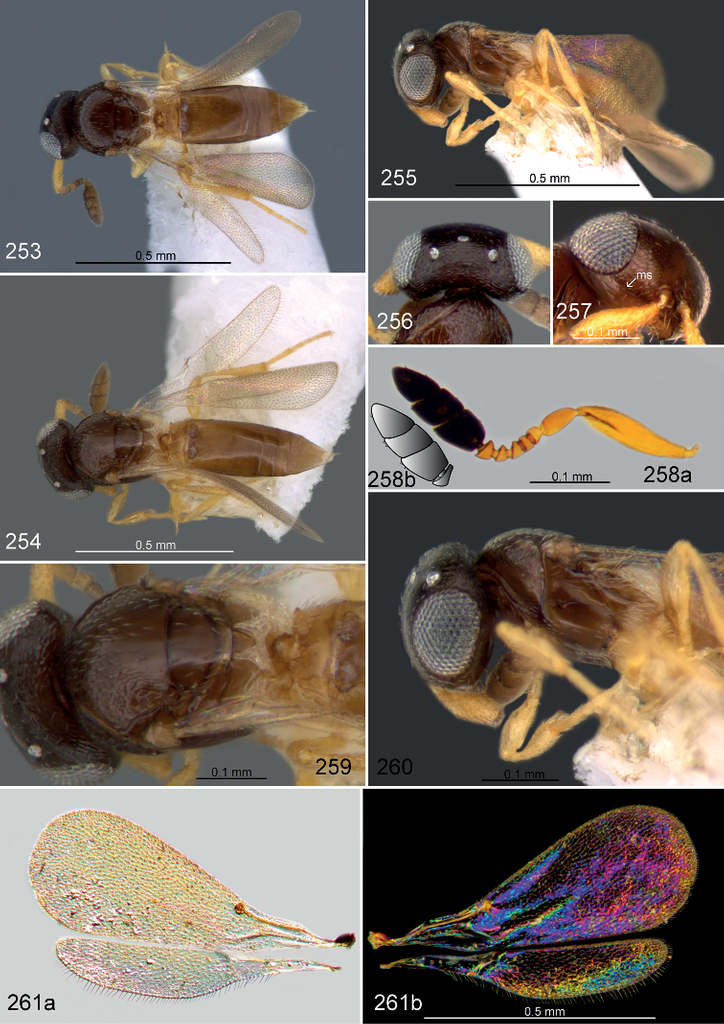
Mesosoma
(Figs
Metasoma
(Figs
Etymology
This species is named after Pierre Tripotin, collector of the holotype specimen and a tremendous friend of Popovici OA. Noun in the genitive case.
Male. unknown.
Material examined
8♀. South Korea: Holotype 1♀, Gangwon-do, Chuncheon Nam-myeon, Hudong-ri, 34.6422°N, 127.6285°E, 31.vii–16.viii.2003, leg. Tripotin P. (MT) (
Paratypes
: 2♀, South Korea, Gangwon-do, Chuncheon Nam-myeon, Hudong-ri, 34.6422°N, 127.6285°E, 6–31.vii.2003, leg. Tripotin P. (MT) (
Distribution
South Korea (Fig.
Biology
unknown.
Diagnosis
This species can be recognized by the presence of a malar sulcus, the short transepisternal line and T2 which is elongate and longer than wide. All other Palearctic Fidiobia with 10-merous antennae have T2 wider than long and the malar sulcus absent.
Comments
Fidiobia tripotini is the only known species of the genus with a malar sulcus. The malar sulcus is not flanked by striation, a state that is found only in Orwellium Johnson, Masner and Musetti among extant Platygastridae. Other extant platygastrids with a malar sulcus, e.g. Metaclisis Förster, have facial and malar striae.
Fidiobia vanharteni
Fidiobia vanharteni Buhl, 2010b: 306.
Description
Female. Body length: 0.7–0.8 mm. Colour of body: melanic (Figs
Head
(Figs

Mesosoma
(Figs

Metasoma
(Figs
Male. unknown.
Material examined
18♀. UAE: Holotype ♀, (Figs
Non-type material
1♀, UAE, al-Ajban, N24.6, E55.016, 3.i–18.ii.2007, leg. van Harten A. (LT), (
Yemen: 1♀, Ghail Ba Wazir, N14.77, E49.37, ?.xi–xii.2002, van Harten A. and Hubaishan M. (MT) (
Distribution
Yemen, UAE (Fig.
Diagnosis
Fidiobia vanharteni is relative morphologically similar to F. hofferi because of fore wings with short, hardly visible marginal fringe, presence of epitorular carina and metascutellum visible in dorsal view, but it can be recognized by its light coloration, faintly banded fore wing (uniformly hyaline in F. hofferi), and OOL slightly shorter or equal to the ocellar diameter (OOL is equal to about 2 OD in F. hofferi).
Fidiobia vladlubomiri , sp. nov.
Description
Female. Body length: 0.8–1.0 mm. Colour of body: melanic (Fig.
Head
(Figs

Fidiobia vladlubomiri: 273 ♀, habitus, dorsal view (OPPC0331) 274 ♂, habitus, dorsal view (OPPC0502) 275 head, dorsal view 276 head, frontal view 277a antenna (♀) (OPPC0313) 277b sensillar formula 278 antenna (♂) (OPPC0502) 279 mesosoma, dorsal view 280 mesosoma, lateral view 281a wings (OPPC0313) 281b WIP.
Mesosoma
(Figs

Metasoma
(Fig.

Male (Fig.

Etymology

Material examined
13♀ and 1♂. South Korea: Holotype 1♀, Chungnam, Daejeon-si, Wadong, 36.3601°N, 127.2345°E, 19.vi–24.vii.2007, leg. Tripotin P. (MT) (OPPC0047).

Paratypes
: South Korea: 1♀, Chungnam, Daejeon-si, Wadong, 36.3601°N, 127.2345°E, 24.vii–21.viii.2007, leg. Tripotin P. (MT) (OPPC0524); 1♀, Chungnam, Daejeon-si, Wadong, 36.3601°N, 127.2345°E, 24.vii–21.viii.2007, leg. Tripotin P. (MT) (OPPC0331); 1♀, Chungnam, Daejeon-si, Wadong, 36.3601°N, 127.2345°E, 24.vii–21.viii.2007, leg. Tripotin P. (MT) (OPPC0313); 1♀, Chungnam, Daejeon-si, Wadong, 36.3601°N, 127.2345°E, 24.vii–21.viii.2007, leg. Tripotin P. (MT) (OPPC0332); 1♀, Chungnam, Daejeon-si, Wadong, 36.3601°N, 127.2345°E, 25.ix–17.xi.2007, leg. Tripotin P. (MT) (OPPC0543); 1♀, Gangwon-do, Chuncheon Nam-myeon, Hudong-ri, 34.6422°N, 127.6285°E, 17.viii–5.ix.2003, leg Tripotin P. (MT) (OPPC0734); 1♀, Chungbuk, Okcheon-gun Dongi-myeon, Soesan-li, 36.2764°N, 127.6131°E, 8–23.vii.2004, leg. Tripotin P. (MT) (OPPC0729); 1♀, Chungbuk, Okcheon-gun Dongi-myeon, Soesan-li, 36.2764°N, 127.6131°E, 19–28.vi.2004, leg. Tripotin P. (MT) (OPPC0061); 1♀, Kangwon, Chuncheon, Nam-myeon, Hudong-li, 6–31.vii.2008, leg. Tripotin P., pastured area, trail close to forest edge (MT) (

Laos: 1♀, Houa Phan, Phou Pane Mt., 1480–1510 m, 20°13'09"N, 103°59'54"E, 1–16.vi.2009, primary forest, leg. Kubáň V., (FIT), (

Japan: 1♀, Aichi, Shitara, Uradani, 18–24.vii.1994, leg. Yamagishi K., (YPT, beech forest) (

Distribution

Biology

Diagnosis
Fidiobia vladlubomiri is a distinct species that can be recognized by wide, deeply incised notauli with the lateral margins located medial to the axillular carina, meeting the scutoscutellar sulcus. The transepisternal line is nearly complete and sigmoid in shape. The dorsal mesopleural area has some transverse striae, and between these striae and the transepisternal line there is a large unsculptured area (in F. striatitergitis and F. nipponica this area is transversely striate). The dorsal metapleural area is covered with dense silvery setae that easily distinguish F. vladlubomiri from F. striatitergitis and F. nipponica. The papillary sensillum located at the apex of the distal clavomere (Fig.


Acknowledgements
We thank Dr. Lars Vilhelmsen (
Also, we wish to thank to Dr. Norman Johnson and Dr. Luciana Musetti for the access to vSysLab platform and for the maintaining the internet site “Hymenoptera Online Database” (https://mbd-p.asc.ohio-state.edu/hol/), a very valuable resource for any Platygastroidea student and to Cristina Vasilița and Jessica Awad for their invaluable comments on the manuscript.
This research received support partially from the SYNTHESYS Project (GB-TAF-1303; HU-TAF-6368 and FR-TAF-6379) http://www.synthesys.info/ which is financed by European Community Research Infrastructure Action under the FP7 “Capacities” Program.
Authors are thankful to Romanian Ministry of Research, Innovation and Digitization, within Program 1 – Development of the national RD system, Subprogram 1.2 – Institutional Performance – RDI excellence funding projects, Contract no.11PFE/30.12.2021, for financial support”. Elijah Talamas was supported by the Florida Department of Agriculture and Consumer Services, Division of Plant Industry.
References
- Alekseyev VN (1979) A new species of the genus Isolia (Hymenoptera, Platygastridae) from Central Asia. Zoologicheskii Zhurnal 58: 763–764. https://doi.org/10.5281/zenodo.23374
- Asadi-Farfar M, Karimpour Y, Lotfalizadeh H, Caleca V (2020) New records of Sceliotrachelinae (Hymenoptera: Platygastridae) in Iran. Journal of Insect Biodiversity and Systematics 6(2): 125–133. https://doi.org/10.52547/jibs.6.2.125
- Ashmead WH (1894) A new genus and species of Proctotrypidae and a new species of Brachysticha, bred by Prof. F. M. Webster. Journal of the Cincinnati Society of Natural History 17: 170–172. https://doi.org/10.5281/zenodo.23424
- Brèthes J (1916) Hyménoptères parasites de l’Amérique méridionale. Anales del Museo Nacional de Historia Natural de Buenos Aires 27: 401–430. https://doi.org/10.5281/zenodo.23610
- Brown BV (1993) A further chemical alternative to critical-point-drying for preparing small (or large) flies. Fly Times 11: 10.
- Brues CT (1909) Notes and descriptions of North American parasitic Hymenoptera. VII. Bulletin of the Wisconsin Natural History Society 7: 154–163. https://doi.org/10.5281/zenodo.23625
- Buhl PN (1998) On some new or little-known NW European species of Platygastridae (Hymenoptera, Proctotrupoidea). Fragmenta Entomologica, Roma 30: 295–334.
- Buhl PN (1999a) A synopsis of the Platygastridae of Fennoscandia and Denmark (Hymenoptera, Platygastroidea). Entomofauna, Zeitschrift für Entomologie 20(3): 17–52.
- Buhl PN (1999b) Platygastridae (Hymenoptera) species of a Juniperus thurifera L. forest of Los Monegros region (Zaragoza, Spain). Zapateri, Revista aragonesa de Entomologia 8: 11–42.
- Buhl PN (2001) Taxonomical notes on Platygastridae (Hymenoptera, Platygastroidea). Entomofauna, Zeitschrift für Entomologie 22: 17–40.
- Buhl PN (2002) On a Baltic amber collection of Platygastridae and Diapriidae (Hymenoptera). Entomologiske Meddelelser 70: 57–61.
- Buhl PN, O’Connor JP, Ashe P (2009) New species of Platygastridae (Hym., Platygastroidea) from Sulawesi. Entomologists Monthly Magazine 145: 87–96.
- Buhl PN (2010a) Platygastridae from the Udzungwa Mountains, Tanzania (Hymenoptera, Platygastroidea). Journal of Afrotropical Zoology 6: 29–46.
- Buhl PN (2010b) Order Hymenoptera, family Platygastridae (part 2). Arthropod Fauna of the United Arab Emirates 3: 306–318.
- Buhl PN (2011) New Neotropical species of Platygastrinae and Sceliotrachelinae (Hymenoptera: Platygastridae), with keys to species of the larger genera, some redescriptions and a checklist. Folia Heyrovskyana 19(1–4): 25–128.
- Buhl PN (2014) New species and new records of Platygastrinae and Sceliotrachelinae from Togo (Hymenoptera: Platygastridae). African Invertebrates 55(1): 67–117. https://doi.org/10.5733/afin.055.0106
- Buhl PN, Creutzburg F, Müller J, Müller J (2016) Checkliste der Platygastridae (Hymenoptera, Proctotrupoidea) Thüringens. Check-Listen Thüringer Insekten und Spinnentiere 24: 5–9.
- Crawford JC (1916) Some new American Hymenoptera. Insecutor Inscitiae Menstruus 4: 135–144.
- Debauche HR (1947) Scelionidae de la faune belge (Hymenoptera Parasitica). Bulletin et Annales de la Société Entomologique de Belge 83: 255–285. https://doi.org/10.5281/zenodo.23744
- Eady RD (1968) Some illustrations of microsculpture in the Hymenoptera. Proceedings of the Royal Entomological Society London. Series A. General entomology 43: 66–72. https://doi.org/10.1111/j.1365-3032.1968.tb01029.x
- Evans GA, Peña JE (2005) A new Fidiobia species (Hymenoptera: Platygastridae) reared from eggs of Diaprepes doublierii (Coleoptera: Curculionidae) from Dominica. Florida Entomologist 97: 61–66. https://doi.org/10.1653/0015-4040(2005)088[0061:ANFSHP]2.0.CO;2
- Fabritius K (1974) Sceliotrachelinae Brues, 1908 (Hymenoptera, Platygasteridae) neu fur die Fauna Rumaniens. Travaux du Muséum National d’Histoire Naturelle “Grigore Antipa” 15: 293–295.
- Fouts RM (1924) Revision of the North American wasps of the subfamily Platygasterinae. Proceedings of the United States National Museum 63(15): 1–145. https://doi.org/10.5479/si.00963801.63-2484.1
- Fusu L (2017) An integrative taxonomic study of European Eupelmus (Macroneura) (Hymenoptera: Chalcidoidea: Eupelmidae), with a molecular and cytogenetic analysis of Eupelmus (Macroneura) vesicularis: several species hiding under one name for 240 years. Zoological Journal of the Linnean Society 181(3): 519–603. https://doi.org/10.1093/zoolinnean/zlw021
- Fusu L, Polaszek A (2017) Description, DNA barcoding and phylogenetic placement of a remarkable new species of Eopelma (Hymenoptera: Eupelmidae) from Borneo. Zootaxa 4263: 557–566. https://doi.org/10.11646/zootaxa.4263.3.7
- Ghesquière J (1948) Note critique sur les genres Xestonotus Foertster et Rosneta Brues (Hymenoptera, Proctotrupoidea, Platygasteridae). Bulletin et Annales de la Société Royale d’entomologie de Belgique 84: 41–45. https://doi.org/10.5281/zenodo.24398
- Harris RA (1979) A glossary of surface sculpturing. Occasional Papers in Entomology 28: 1–32. https://doi.org/10.5281/zenodo.26215
- Jansson A (1956) Studier over svenska proctotrupider. 10. Fidiobia tatrae Szel. (Platygasteridae, Inostemmatinae, adhuc falso Platygasterinae), en art med utbredd foerekomst i Sverige. Entomologisk Tidskrift 77: 87–90.
- Kamalanathan V, Buhl PN, Rajmohana K, Mohanraj P (2012) Three new species of Sceliotrachelinae (Hymenoptera: Platygastroidea: Platygastridae) from South India. Entomofauna, Zeitschrift für Entomologie 33(33): 469–480.
- Kamalanathan V, Popovici OA, Buhl PN, Mohanraj P (2018) Revision of Indian Species of Fidiobia Ashmead (Platygastridae: Sceliotrachelinae). Annales Zoologici 68(3): 553–600. https://doi.org/10.3161/00034541ANZ2018.68.3.013
- Kamalanathan V, Buhl PN, Mohanraj P (2019) Review of the genus Isolia Förster (Platygastroidea: Platygastridae: Sceliotrachelinae) with description of two new species from India. Zootaxa 4565(4): 451–474. https://doi.org/10.11646/zootaxa.4565.4.1
- Kieffer JJ (1921) Un platygasterine parasite d’un cynipide. Brotéria 19: 68–69. https://doi.org/10.5281/zenodo.24387
- Kieffer JJ (1926) Scelionidae. Das Tierreich. Vol. 48. Berlin: Walter de Gruyter & Co, 885 pp.
- Koponen M, Huggert L (1982) Platygastridae (Hymenoptera, Proctotrupoidea) from Finland. Notulae Entomologicae 62: 51–59. https://doi.org/10.5281/zenodo.24456
- Kozlov MA (1971) Proctotrupoids (Hymenoptera, Proctotrupoidea) of the USSR. Trudy Vsesoyuznogo Entomologicheskogo Obshchestva 54: 3–67. https://doi.org/10.5281/zenodo.23678
- Kozlov MA (1978) Superfamily Proctotrupoidea. In: Medvedev GS (Ed.) Determination of insects of the European portion of the USSR. Vol. 3, part. 2. Nauka, Leningrad (Russia), 538–664.
- Kozlov MA (1987) Superfamily Proctotrupoidea (Proctotrupoids). In: Medvedev GS (Ed.) Keys to the insects of the European part of the USSR. Vol. 3 Hymenoptera, part. 2. Amerind Publishing Co. Pvt. Ltd., New Delhi, 983–1212.
- Lahey Z, van Noort S, Polaszek A, Masner L, Johnson NF (2019) Revision of the Afrotropical genus Pulchrisolia Szabó (Hymenoptera, Platygastridae, Sceliotrachelinae). In: Talamas E (Ed.) Advances in the Systematics of Platygastroidea II. Journal of Hymenoptera Research 73: 39–71. https://doi.org/10.3897/jhr.73.33876
- Lahey Z, Musetti L, Masner L, Johnson NF (2021) Revision of Phoenoteleia Kieffer (Hymenoptera, Scelionidae, Scelioninae). In: Lahey Z, Talamas E (Eds) Advances in the Systematics of Platygastroidea III. Journal of Hymenoptera Research 87: 575–611. https://doi.org/10.3897/jhr.87.59794
- Lemarie J (1958) Beitrag zur Kenntnis der Parasiten der Kiefernknospentriebmotte Exoteleia (Heringia) dodecella L. Teil 1. Chalcidoidea, Proctotrupoidea, Bethyloidea. Folia Zoologica 7(3): 221–230.
- Lemarie J (1959) Erster Beitrag zur Kenntnis der Parasiten von Exoteleia dodecella L. (Hym. Chalcidoidea, Proctotrupoidea, Bethyloidea). Beiträge zur Entomologie 9: 805–809.
- Lemarie J (1960) Beitrag zur Kenntnis der Parasiten der Kiefernknospentriebmotte Exoteleia dodecella L. III. (Chalcidoidea, Proctotrupoidea). Folia Zoologica 9(3): 235–236.
- Lemarie J (1961) Beitrag zur Kenntnis der Parasiten der Kiefernknospentriebmotte Exoteleia dodecella L. Teil 4. Ichneumonidae, Braconidae, Chalcidoidea, Bethyloidea. Folia Zoologica 10(2): 119–126.
- Loiacono MS (1982) Un nuevo platygatrido (Hymenoptera- Platygastridae) criado de huevos de Naupactus xanthographus Germ. (Coleoptera-Curculionidae). Revista de la Sociedad de Entomologia de Argentina 41(1–4): 85–88.
- Maneval H (1940) Fam. XVII. Proctotrypides. Pages 93–118 in Perrier R. La Faune de la France en tableaux synoptiques illustrés. Tome VII. Hyménoptères par Lucien Berland avec la collaboration de MM. Raymond Benoit, Francis Bernard, Henri Maneval. Paris, 213 pp.
- Masner L, Huggert L (1989) World review and keys to genera of the subfamily Inostemmatinae with reassignment of the taxa to the Platygastrinae and Sceliotrachelinae (Hymenoptera: Platygastridae). Memoirs of the Entomological Society of Canada 147: 1–214. https://doi.org/10.4039/entm121147fv
- Masner L, Muesebeck CFW (1968) The types of Proctotrupoidea (Hymenoptera) in the United States National Museum. Bulletin of the United States National Museum 270: 1–143. https://doi.org/10.5479/si.03629236.270
- Mikó I, Vilhelmsen L, Johnson NF, Masner L, Pénzes Z (2007) Skeletomusculature of Scelionidae (Hymenoptera: Platygastroidea): head and mesosoma. Zootaxa 1571: 1–78. https://doi.org/10.11646/zootaxa.1571.1.1
- Mikó I, Masner L, Deans AR (2010) World revision of Xenomerus Walker (Hymenoptera: Platygastroidea, Platygastridae). Zootaxa 2708: 1–73. https://doi.org/10.11646/zootaxa.2708.1.1
- Muesebeck CFW, Masner L (1967) In: Krombein and Burks (Ed.) Hymenoptera of America north of Mexico. Synoptic Catalog (Agriculture Monograph No. 2). Second supplement. United States Government Printing Office, Washington 584 pp.
- Nixon GEJ (1969) Two new species of Platystasius Nixon with a note on the generic relationship between Platystasius and Fidiobia (Hymenoptera: Platygasteridae). Proceedings of the Entomological Society of Washington 71: 445–449. https://doi.org/10.5281/zenodo.24443
- Notton DG, Popovici OA, van Achterberg C, De Rond J, Burn JT (2014) Parasitoid wasps new to Britain (Hymenoptera: Platygastridae, Eurytomidae, Braconidae & Bethylidae). European Journal of Taxonomy 99: 1–20. https://doi.org/10.5852/ejt.2014.99
- Noyes JS (1982) Collecting and preserving chalcid wasps (Hymenoptera: Chalcidoidea). Journal of Natural History 16: 315–334. https://doi.org/10.1080/00222938200770261
- O’Connor JP, Nash R, Notton DG, Ferguson NDM (2004) A catalogue of the Irish Platygastroidea and Proctotrupoidea (Hymenoptera). Occasional Publication of the Irish Biogeographical Society 7: 1–110.
- Oglobin AA (1944) Two new species of Proctotrupoidea from Iowa (Hymenoptera). Proceedings of the Entomological Society of Washington 46: 155–158.
- QGIS Development Team (2021) QGIS Geographic Information System. Open Source Geospatial Foundation Project. http://qgis.osgeo.org
- Popovici OA, Buhl PN (2010) The West Palearctic species of Fidiobia Ashmead, 1894 (Hymenoptera: Platygastroidea). Journal of Natural History 44(19–20): 1131–1164. https://doi.org/10.1080/00222931003632740
- Popovici OA, Masner L, Polaszek A (2018) A revision of the European species of Baeoneurella Dodd (Hymenoptera: Scelionidae). Journal of Natural History 52(43–44): 2745–2794. https://doi.org/10.1080/00222933.2018.1546913
- Szabó JB (1958) Erganzende Beobachtungen uber die holarktische Gattung Fidiobia Ashmead 1894. Folia Entomologica Hungarica 11: 457–464.
- Szabó JB (1962) Untersuchungen an palaearktischen Proctotrupiden. I–IV. (Hymenoptera). Folia Entomologica Hungarica 15: 221–243.
- Szelényi G (1938) Über paláarktische Scelioniden (Hym., Proct.). Annales Historico-Naturales Musei Nationalis Hungarici 31: 108–128.
- Szelényi G (1941) Neue Gattungen und Arten der palaearktischen Scelioniden (Hym., Proctotrupoidea). Zoologischer Anzeiger 134: 158–168.
- Shevtsova E, Hansson C (2011) Species recognition through wing interference patterns (WIPs) in Achrysocharoides Girault (Hymenoptera, Eulophidae) including two new species. ZooKeys 154: 9–30. https://doi.org/10.3897/zookeys.154.2158
- Shevtsova E, Hansson C, Janzen DH, Kjærandsen J (2011) Stable structural color patterns displayed on transparent insect wings. Proceedings of the National Academy of Sciences, USA 108(2): 668–673. https://doi.org/10.1073/pnas.1017393108
- Talamas EJ, Buffington ML (2015) Fossil Platygastroidea in the National Museum of Natural History, Smithsonian Institution. Journal of Hymenoptera Research 47: 1–52. https://doi.org/10.3897/JHR.47.5730
- Tereshkin AM (1990) A trap for the xylobiontes and theirs parasites. http://tereshkin.info/papers/a-trap-for-the-xylobiontes-and-theirs-parasites-inrussian.html [In Russian]
- van Noort S, Lahey Z, Talamas EJ, Austin AD, Masner L, Polaszek A, Johnson NF (2021) Review of Afrotropical sceliotracheline parasitoid wasps (Hymenoptera, Platygastridae). In: Lahey Z, Talamas E (Eds) Advances in the Systematics of Platygastroidea III. Journal of Hymenoptera Research 87: 115–222. https://doi.org/10.3897/jhr.87.73770
- Varga O (2017) A review of the tribe Pimplini Wesmael, 1845 (Hymenoptera, Ichneumonidae, Pimplinae) from the Carpathians, with new records for Romania and Ukraine. Turkish Journal of Zoology 41: 354–362. https://doi.org/10.3906/zoo-1604-75
- Vlug HJ (1995) Catalogue of the Platygastridae (Platygastroidea) of the world (Insecta: Hymenoptera). Hymenopterorum Catalogus 19: 1–168.
- Yoder MJ, Mikó I, Seltmann KC, Bertone MA, Deans AR (2010) A gross anatomy Ontology for Hymenoptera CS Moreau (Ed.). PLoS ONE 5(12): e15991. https://doi.org/10.1371/journal.pone.0015991